Knowledge Hub
Featured Articles
Latest Articles
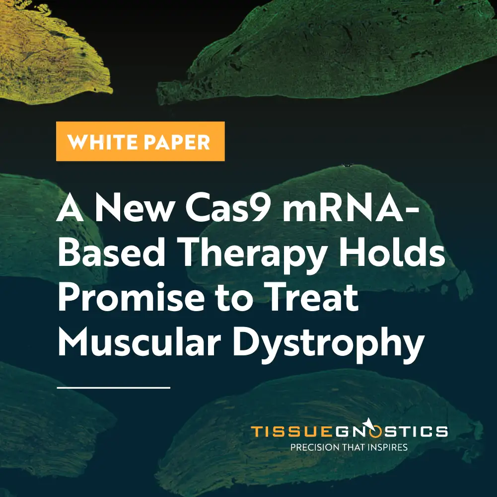
White Paper
13 May, 2026
A New Cas9 mRNA‑Based Therapy Holds Promise to Treat Muscular Dystrophy
Quantitative confocal imaging helps researchers to confirm muscle protein restoration after Cas9 mRNA delivery via targeted lipid nanoparticles.
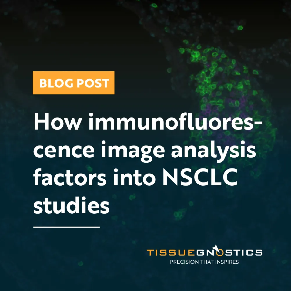
Blog Post
07 Apr, 2026
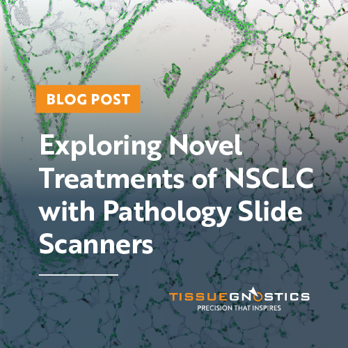
How immunofluorescence image analysis factors into NSCLC studies
It’s crucial to understand how immunofluorescence image analysis factors into NSCLC studies to uncover spatial biomarkers and guide research.
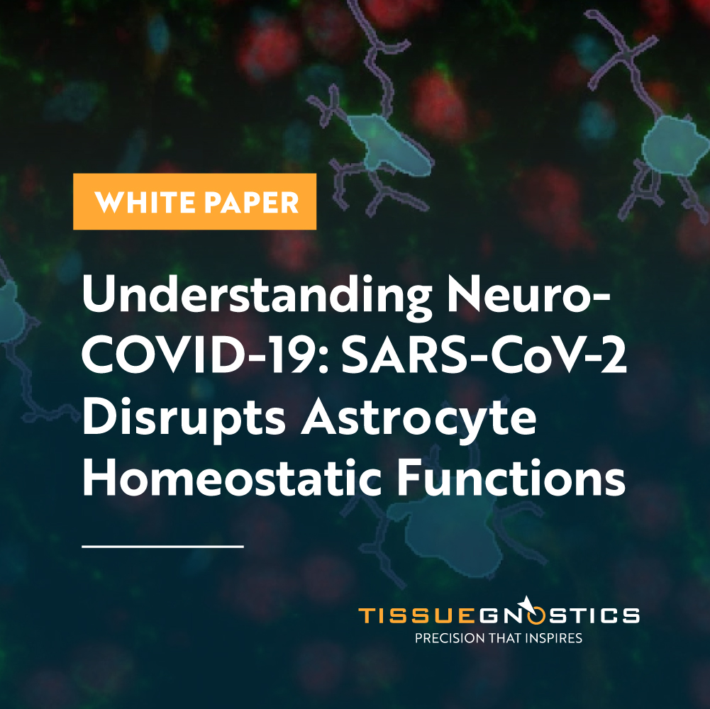
White Paper
30 Mar, 2026
Understanding NeuroCOVID-19: SARS-CoV-2 Disrupts Astrocyte Homeostatic Functions
Quantitative cellular analysis of COVID-19-infected astrocyte cells reveals a more central role of astrocytes in NeuroCOVID-19 and the interruptive impact of SARS-CoV-2 on astrocyte homeostasis capabilities, reframing astrocytes as active protagonists.

Customer Publication
11 Mar, 2026
Spatial Multi-Omics Identifies NAD-Driven Niche in Early Gastric Cancer
A new study reveals how an NAD-dependent immunosuppressive niche drives early gastric cancer development. Using spatial multi-omics and TissueFAXS Spectra with StrataQuest, researchers mapped cell interactions and quantified tumor-promoting fibroblast and epithelial populations.

Press Release
18 Feb, 2026
TissueGnostics Expands Its Presence in Australia: Partnership with Klein Scientific
TissueGnostics and Klein Scientific have entered a partnership to bring advanced microscopy solutions to researchers and institutions across Australia. The collaboration expands TissueGnostics’ reach in the Asia-Pacific region and gives Klein Scientific customers access to tissue cytometry and spatial phenotyping tools.
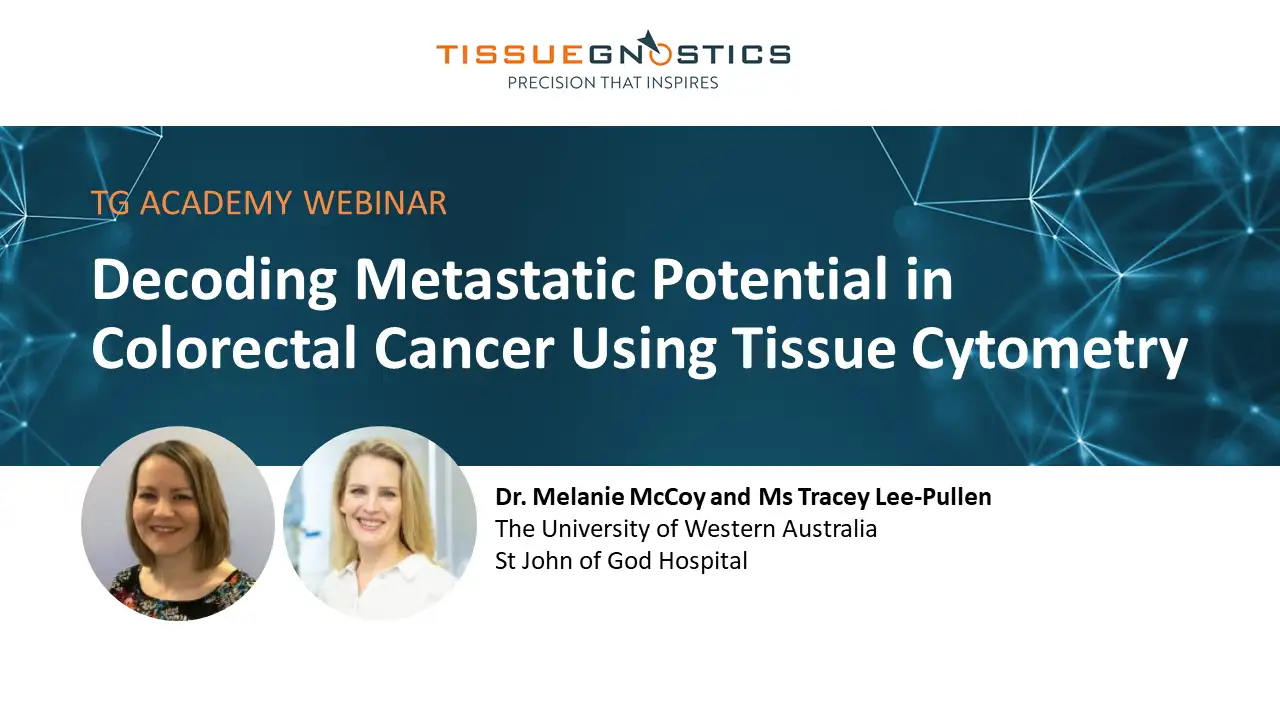
Webinar
20 Jan, 2026
Decoding Metastatic Potential in Colorectal Cancer Using Tissue Cytometry
At ASI 2025, Dr. Melanie McCoy and Ms. Tracey Lee-Pullen presented research on metastatic colorectal cancer, using quantitative tissue cytometry and StrataQuest to study whether the tumor microenvironment predicts metastasis.
Webinar
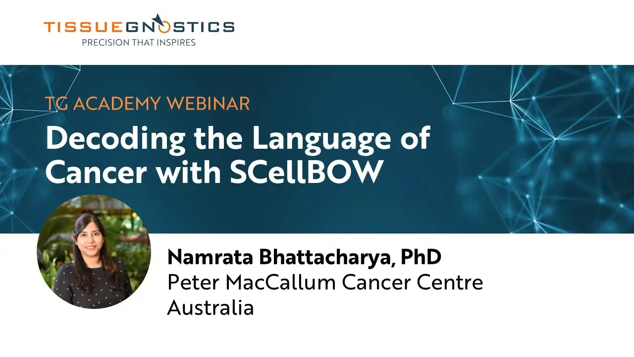
12 Nov, 2025
TG Academy Webinar - Decoding the language of cancer with SCellBOW
Dr. Namrata Bhattacharya, Peter MacCallum Cancer Centre, presents her work on SCellBOW (Single-Cell Bag-of-Words) - a novel computational approach that facilitates effective identification and high-quality visualization of single-cell subpopulations.