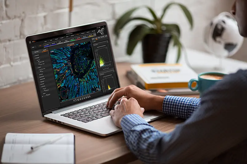
Solutions from Slide to Insight
From whole-slide scanning to AI-powered tissue analysis, TissueGnostics offers modular systems and intelligent products that grow with your research.


Customizable and Flexible Solutions
Built for your research needs.
Every research project is different. That’s why our systems are designed to adapt—offering a modular architecture that supports a wide range of imaging modalities, experimental designs, and future upgrades. Whether you're starting with standard configurations or planning something more specialized, you’ll have the flexibility to shape the system around your scientific objectives.
What sets TissueGnostics apart is not just the hardware—it’s the partnership. Our multidisciplinary team works closely with you to ensure your setup evolves alongside your research, providing the insight and customization options needed to turn complex ideas into actionable results.
Built Around Your Science
Flexible systems, intuitive workflows, and expert support—everything you need to drive your research forward with confidence.
Solutions for Every Research Area
Whether you're working in immunology, oncology, neuroscience, or beyond—our broad portfolio of scanning and analysis tools ensures you’ll find the right fit for your scientific goals.
Seamless Slide-to-Insight Workflow
Move from slide scanning to image analysis without switching platforms. Our integrated workflow eliminates bottlenecks, reduces manual steps, and accelerates your path from raw data to publication-ready results.
Customizable to Your Needs
Maintain full control over your research. From microscopy configuration to software parameters, our solutions are built to be highly customizable—empowering users to fine-tune every step to match their experimental needs.
Reliable Support at Every Stage
From initial setup to advanced troubleshooting, our expert team is here to support you every step of the way—before, during, and after your project begins.
Image Analysis Solutions
Build custom pipelines with StrataQuest or jump-start your workflow with preconfigured apps from the AppCenter.
StrataQuest Image Analysis
Turns imaging data into publication-ready results
- Customizable pipelines for basic & advanced image analysis
- AI-powered nuclei segmentation and tissue classification
- Spatial analysis tools (neighborhood, distance, clustering)
- Integrated data mining: t-SNE, UMAP, SONG, and more
- Prebuilt analysis workflows for oncology, neuroscience & more
- Open data format support and integration with TissueFAXS systems
StrataQuest AppCenter
Library of expert-built analysis pipelines
- Preconfigured analysis pipelines for a number of research areas
- Oncology, immunology, neuroscience, bone research, cardiology, dermatology & more
- Customize pre-built apps or build your own with modular analysis components
- Standardize analysis across users, sites, and studies
- Visualize results with layered outputs, graphs, and overlays
Flexible Imaging Platforms
Compare our scalable TissueFAXS platform with the all-in-one Colubris system to find the best fit for your imaging and analysis needs.

TissueFAXS Imaging Platform
Modular systems built to grow with your research
- Brightfield, fluorescence, multispectral, confocal and high-throughput imaging
- Component-level flexibility: upgrade optics, lightsources, and cameras as needed
- Available in upright and inverted configurations
- Optional darkfield, phase contrast, DIC, and polarization upgrades
- Compatible with slides, well plates, chamber slides, and Petri dishes
- Seamless integration with StrataQuest image analysis
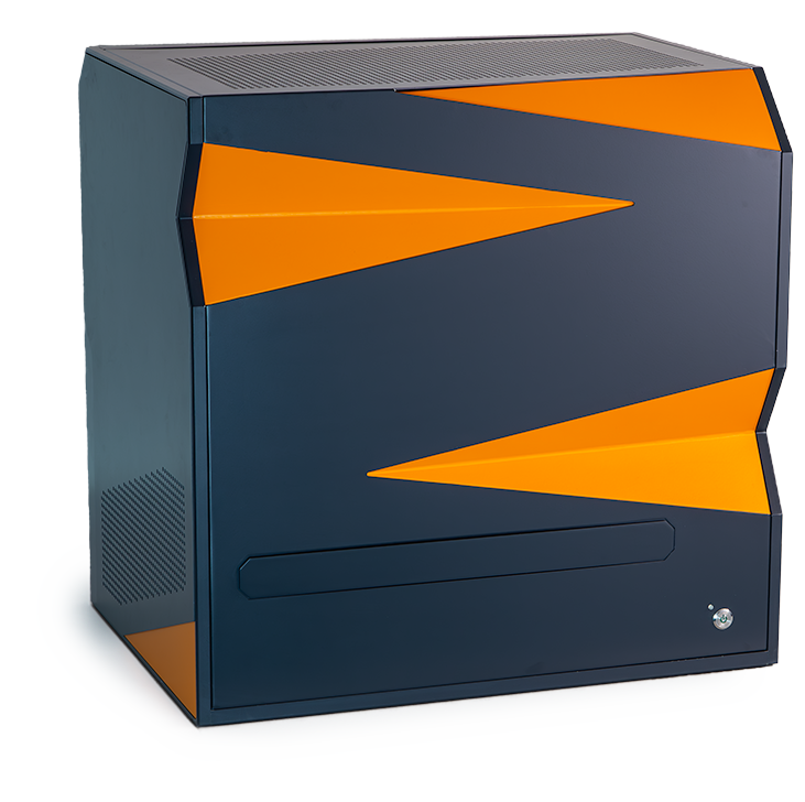
Colubris - Boxed system
All-in-one acquisition and analysis platform
- Combines scanning, analysis, and figure generation in a single, integrated system
- Real-time multitasking with GPU-accelerated image processing
- Customizable, on-the-fly workflows for maximum flexibility
- Explore, structure, and interpret large quantities of imaging data and analysis results
- Share and interactively review data, control the acquisition process and launch analyses remotely
- Ideal for small labs, satellites, and decentralized workflows
The TissueFAXS Family
Four main configurations for different imaging workflows—all part of one scalable, modular platform.
Multispectral Fluorescence Imaging
TissueFAXS Spectra & Spectra SL
- Detect more markers in a single imaging round with LCTF and lamda stacking technology
- Reduce spectral overlap and bleed-through with spectral unmixing
- Precise unmixing with an extensive reference spectra database
- Ideal for immune profiling, tumor microenvironment studies, and spatial biology
upright
multispectral-imaging
high-throughput imaging
Brightfield & Fluorescence Imaging
TissueFAXS Plus and i Plus*
- Whole-slide imaging in brightfield and fluorescence
- Upright or inverted configurations for tissue and cell culture imaging
- Compatible with oversized slides, well plates, and chamber slides*
- Upgrade-ready for confocal, multispectral, or slide loading
- Designed for research labs needing a flexible, expandable imaging platform
upright
inverted
brightfield
fluorescence

High-Speed Confocal Imaging
TissueFAXS Q, iQ & SLQ
- High-speed spinning disk confocal for fast z-stack acquisition
- Capture crisp, high-resolution images with optical sectioning
- Reduced photobleaching and phototoxicity
- Excellent for thicker samples or subcellular localization
- Used in neuroscience, co-localization, and high-detail tissue studies
upright
inverted
high-speed confocal imaging
high-throughput imaging
High-Throughput Whole-Slide Imaging
TissueFAXS SL
- Automate batch imaging with up 120-slides per run
- Scan mixed sample types and staining protocols in one batch
- Combine with Multispectral, or Confocal systems
- Ideal for core facilities and large-scale studies
upright
high-speed confocal imaging
multispectral-imaging
high-throughput imaging
TissueFAXS Imaging and Viewing Software

TissueFAXS Viewer
Free. Fast. Made for Collaboration.
TissueFAXS Viewer is a free, standalone desktop application for exploring high-resolution virtual slides generated by TissueFAXS systems. It allows researchers and collaborators to navigate brightfield, fluorescence, or multispectral scans, view annotations, and assess results without needing the full imaging setup. Lightweight and easy to use, it’s the ideal tool for sharing and reviewing data across teams.
annotations
standalone
slide viewing

TissueFaxs Imaging Software
For routine workflows and complex imaging tasks.
TissueFAXS features powerful imaging software designed to support both routine and advanced workflows. Users have full access to all scanning parameters allowing protocols to be precisely tailored and saved as reusable profiles for consistent results. Smart autofocus and Z-stacking with a Z-projection (extended focus) algorithm, improve clarity in thick or uneven samples, resulting in high-quality seamless images.
brightfield
fluorescence
high-speed confocal imaging
multispectral-imaging
Need Help Finding the Right Solution?
Schedule a consultation with our experts and get personalized guidance based on your research goals and workflow needs.
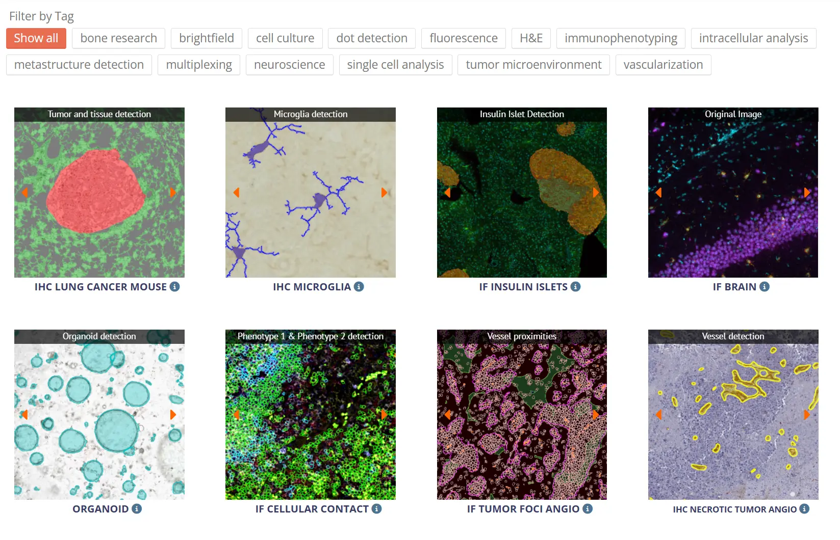
Explore our Products in Action
Our knowledge base provides valuable resources where Tissuegnostics' products were employed to push science forward - white papers, application notes and webinars.
Couldn't find the answer to your question? Need assistance?
Submit an inquiry and one of our TissueGnostics Experts will get back to you
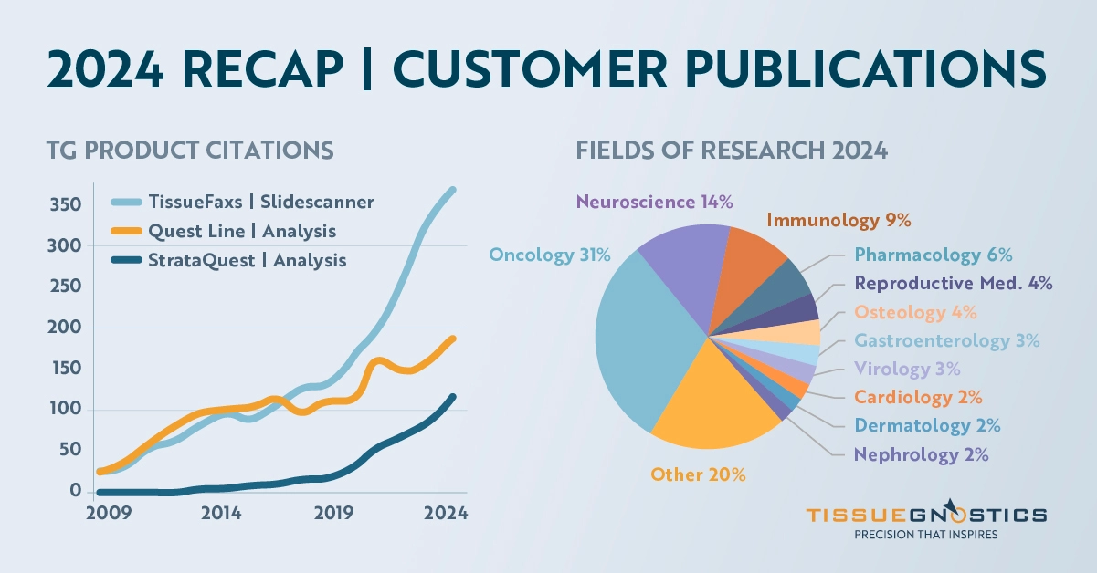
Trusted by Thousands of Researchers
Over 3,200 publications using TG solutions
Researchers worldwide rely on TG imaging and analysis platforms to generate high-quality, reproducible data across a wide range of fields—from oncology and immunology to neuroscience and regenerative medicine.
