


TISSUE CYTOMETRY TECHNOLOGY
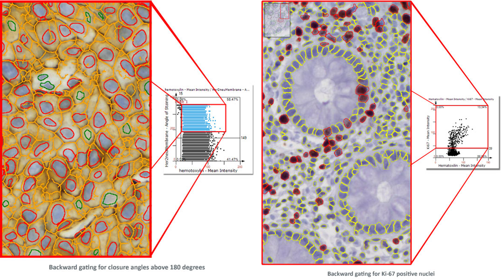
TG introduced Tissue Cytometry technology into image analysis software more than a decade ago with TissueQuest software in its first version. While, at a first look, it may appear to be just another form of displaying data, there is much more to it. Combined with segmentation images it is also a simple and intuitive tool to visually validate results. TG calls this „Backward Gating“ and „Forward Gating“.
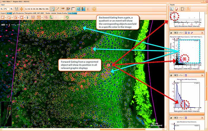
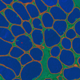
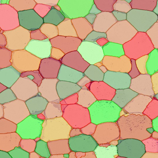
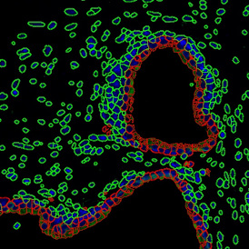
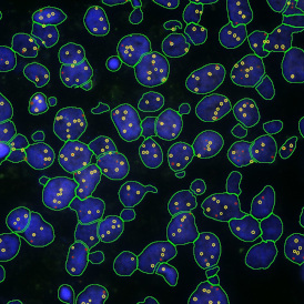
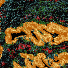
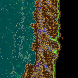
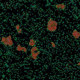
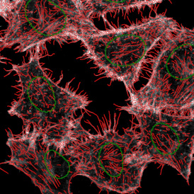
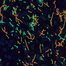
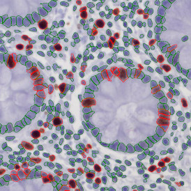
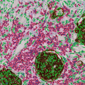
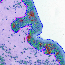
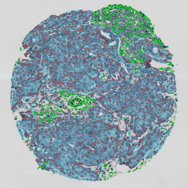
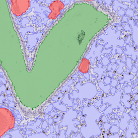
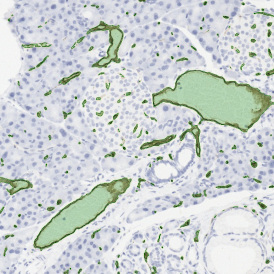
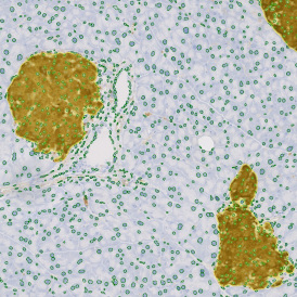
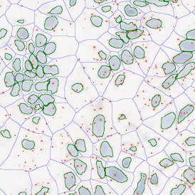
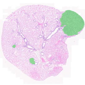
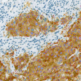
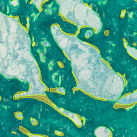
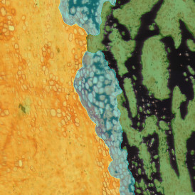
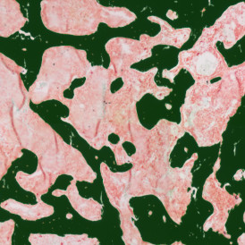
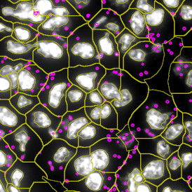
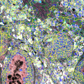
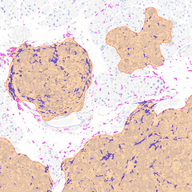
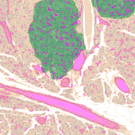
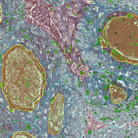
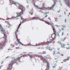
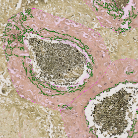
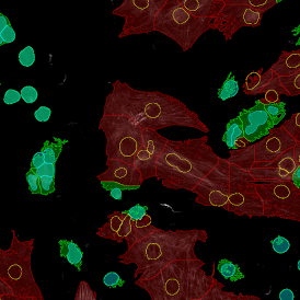
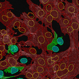
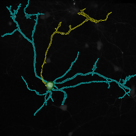
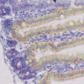
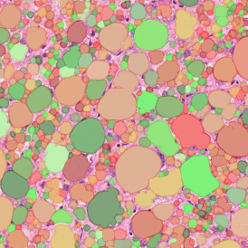
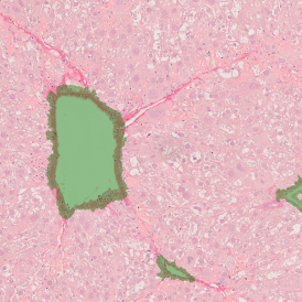
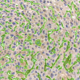
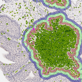
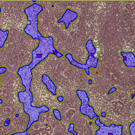
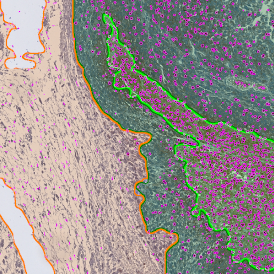
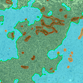
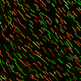
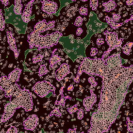
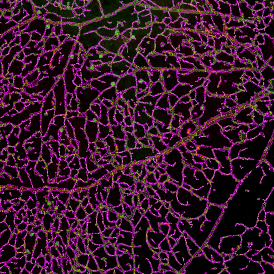
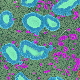
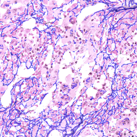
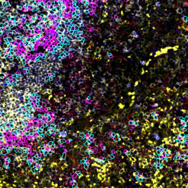
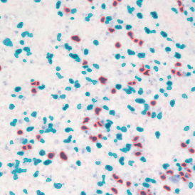
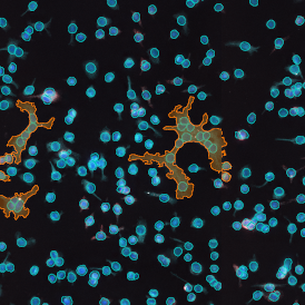
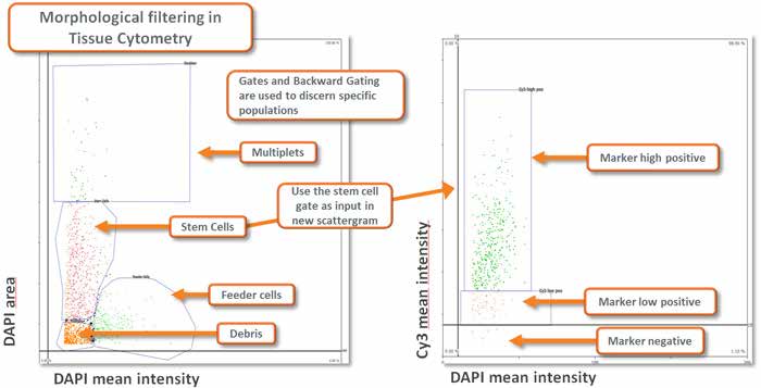
Morphological filtering using Tissue Cytometry technology combined with appropriate planning of the analysis provides a highly flexible and capable instrument for extracting specific data from a sample (see image below).

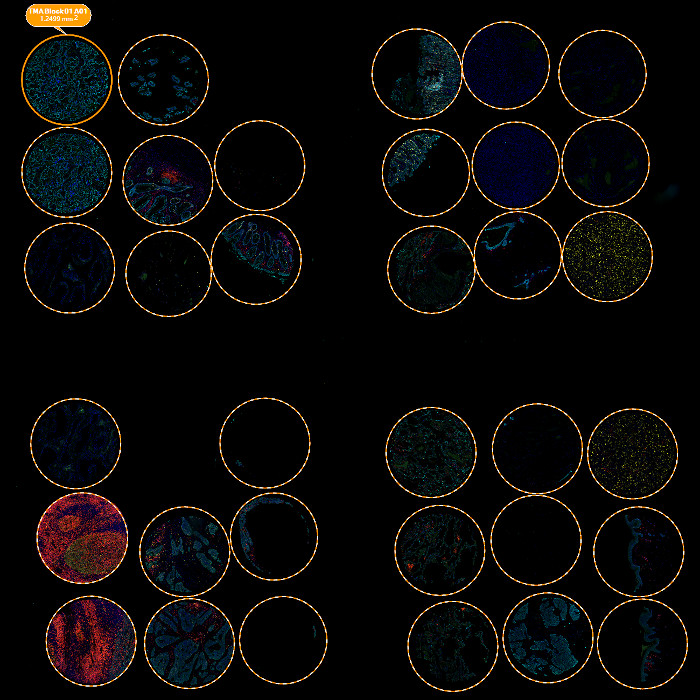
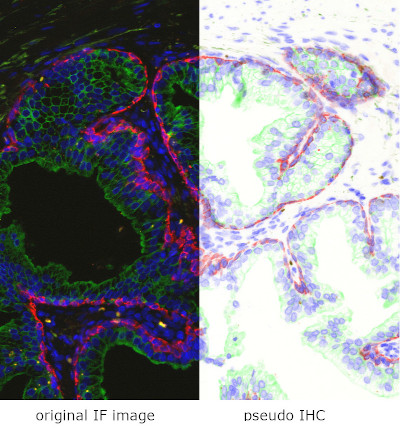 ation of images, handling of TMAs, and the user interface. Here are some highlights: