30
AUG
30. August 2023
What is Multiplexing & How does it Work?
Multiplexing combines multiple measurements in a single experiment. In tissue cytometry, multiplexing involves using multiple fluorophores that emit at different wavelengths or even multiple de-/staining rounds and thereby increase the number of markers that can be visualized. Multiplexing allows for the rapid acquisition of more data per sample.
Analytical assays have become widely used in biomedical research and medical diagnosis. The detection and quantification of biomarkers/specific cell populations is invaluable as a part of a combined approach with tissue analysis in clinics and research.1 As many microscopy measurements can be time-consuming in terms of scan lengths or samples may require involved and lengthy preparation, maximizing the information from a single experiment is essential, and this is where multiplexing can help.
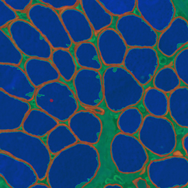
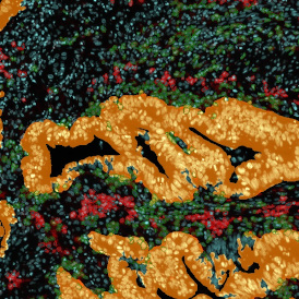
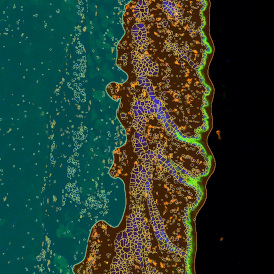
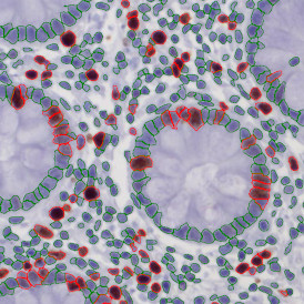
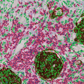
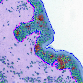
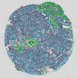
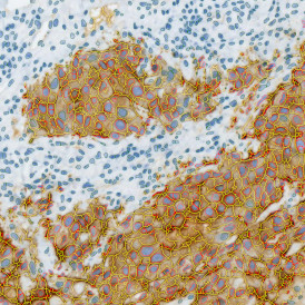
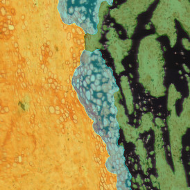
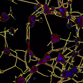
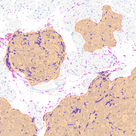
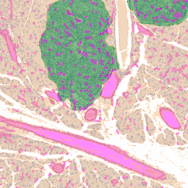
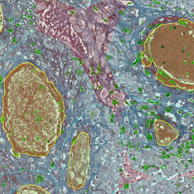
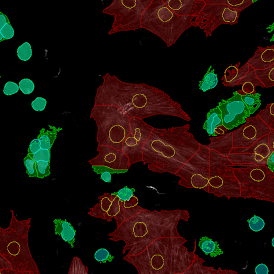
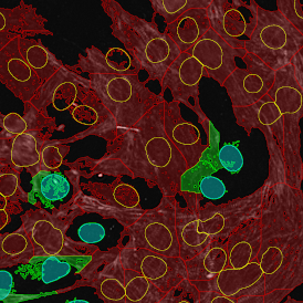
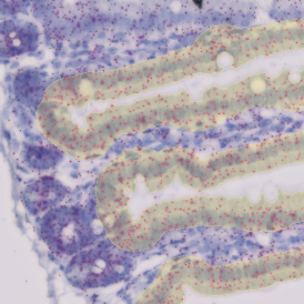
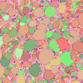
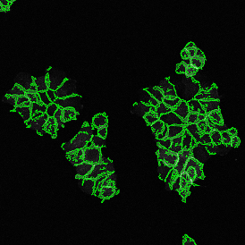
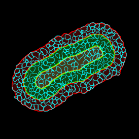
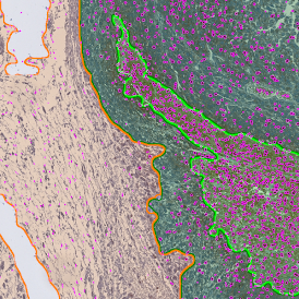
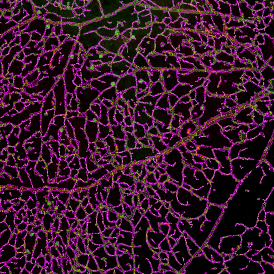
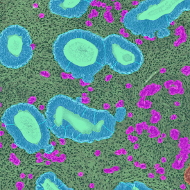
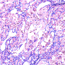
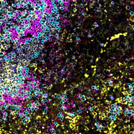
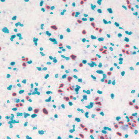
Multiplexing in cancer research merges advanced staining methods with high-tech imaging and analysis. Traditional techniques like immunohistochemistry (IHC) and immunofluorescence (IF) can only detect limited markers in a tissue. However, multiplexing, using special staining protocols, allows simultaneous visualization of multiple markers, producing more information-rich images.
Several established protocols are now available for multiplex imaging based on IHC approaches or IF. Despite multiplexing being a relatively recent development for understanding complex tumor microenvironments, new protocol development is happening rapidly to fully exploit the opportunities in immunology offered by multiplexing.3 Companies such as TissueGnostics, who are experts in bioimaging applications and image analysis, also now have dedicated hardware platforms designed with multiplexing experiments as their focus.
How does it Work?
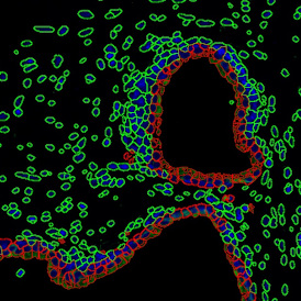
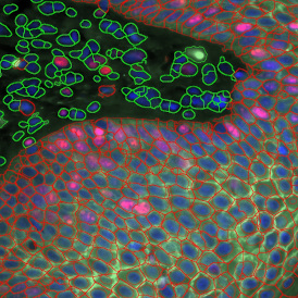
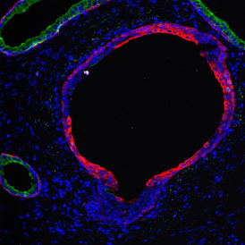
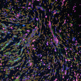
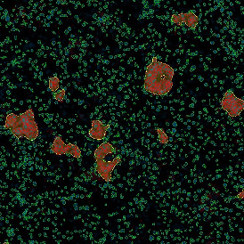
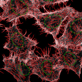
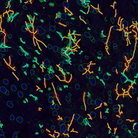
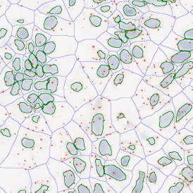
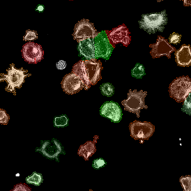
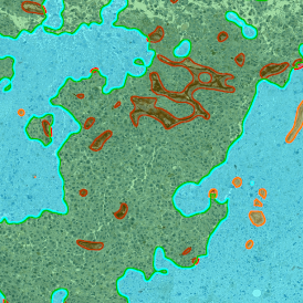
Multiplexing is a powerful technique used in tissue cytometry that allows for the simultaneous analysis of multiple markers within a single tissue sample. This is particularly important in the study of complex biological systems, such as the tumor microenvironment, where numerous cell types and signaling molecules interact.
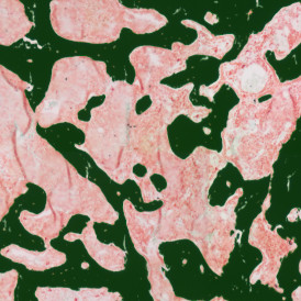
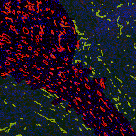
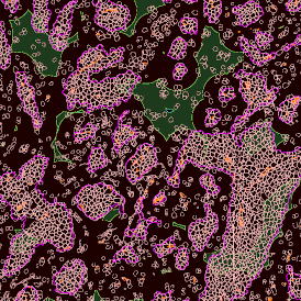
There are two main types of multiplexing methods: immunohistochemistry (IHC)-based and immunofluorescence (IF)-based. Several IHC-based multiplexing methods use the chemical property of alcohol solubility of the peroxidase substrate 3-amino-9-ethylcarbazole (AEC). After image acquisition, the AEC is removed with an organic solvent-based destaining buffer, and the tissue is restained with new antibodies targeting other markers of interest.
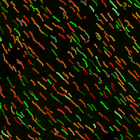
IF-based multiplexing methods, on the other hand, are faster and more effective than IHC-based methods, as they allow for more than one marker to be stained simultaneously in each staining round. Techniques such as “TSA Opal multiplex immunohistochemistry” (Opal mIHC), “in silico multiplexing workflow”, “tissue-based cyclic immunofluorescence” (t-Cycif), and DNA barcoding-based techniques like “CO detection by InDEXing” (CODEX) fall under this category.
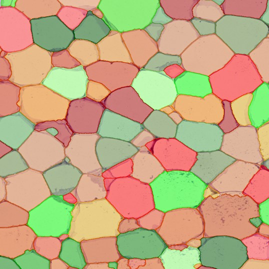
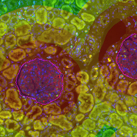
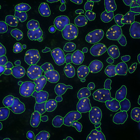
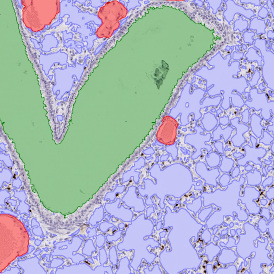
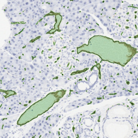
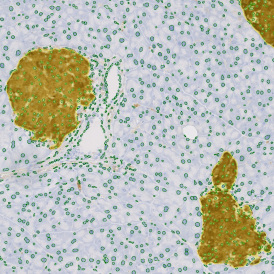
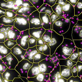
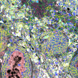
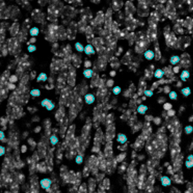
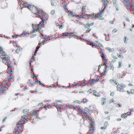
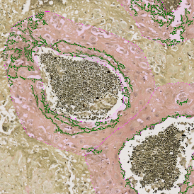
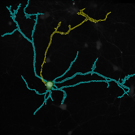
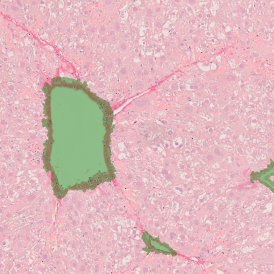
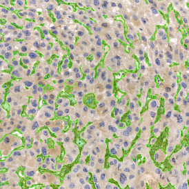
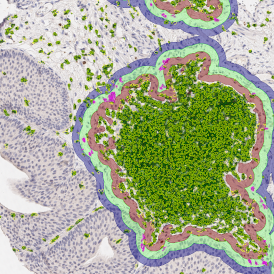
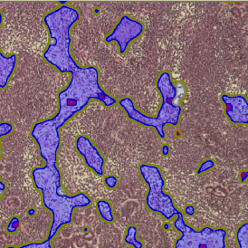
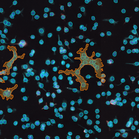
The enhanced number of stained markers offered by multiplexing also increases the necessity for advanced imaging and analysis platforms. These platforms provide fully automated acquisition of the stained tissue sections and computer-assisted high-content phenotypic analysis. The integration of artificial intelligence algorithms for pattern recognition into the image analysis process has been a significant advancement, enabling automatic detection of objects such as nuclei and specific structures within an entire digitized slide.
Data Analysis
Multiplexing has the opportunity to dramatically improve workflows, reduce sample consumption and drastically increase the number of detected markers. However, the wealth of information in multiplex images means sophisticated data analysis methods which are often required to fully capitalize on the opportunities offered.
Contact TissueGnostics today to find out how our specially designed data analysis suites and instrumentation can help you realize the efficiency improvements of multiplex imaging in combination with automation in your laboratory.
References and Further Reading
- Mulrane, L., Rexhepaj, E., Penney, S., Callanan, J. J., Mulrane, L., Rexhepaj, E., & Penney, S. (2014). Automated image analysis in histopathology : a valuable tool in medical diagnostics. Expert Review of Molecule Diagnostics, 8(6), 707–725. https://doi.org/10.1586/14737159.8.6.707
- Mungenast, F., Fernando, A., Nica, R., Boghiu, B., Lungu, B., Batra, J., & Ecker, R. C. (2021). Next-Generation Digital Histopathology of the Tumor Microenvironment. Genes, 12, 538. https://doi.org/10.3390/genes12040538
- Thurin, M., Cesano, A., & Editors, F. M. M. (2020). Biomarkers for Immunotherapy of Cancer. Spinger Protocols. https://link.springer.com/protocol/10.1007/978-1-4939-9773-2_22