Latest articles in

Webinar
26 May, 2025
Exploring Immune Cell Interactions and Tissue Regeneration through Imaging
Dr. Uwe Ritter, LIT, reports on studies of Tregs in wound healing using 3D skin models and CD8+ T cells in tissue regeneration. He further describes the development of a co-cultured organoid detection pipeline published recently.
Customer Publication
13 May, 2025
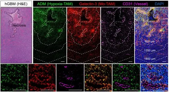
Identification of hypoxic macrophages in glioblastoma with therapeutic potential
Wang et al. used spatial transcriptomics to characterize subsets of tumor-associated macrophages (TAMs) known as monocyte-derived TAMs. They identified a cluster of hypoxia-TAM oversecreting adrenomedullin with multiplex immunostaining imaged with TissueFAXS Spectra, and StrataQuest for marker identification.
Webinar
30 Apr, 2025
Unlocking the Lymphoid Structures with Germinal Centers
Dr. Diana Mechtcheriakova, Medical University of Vienna, presents research on lymphoid structures in systemic immunity and anti-cancer responses, with applications in colorectal cancer.
White Paper
23 Apr, 2025
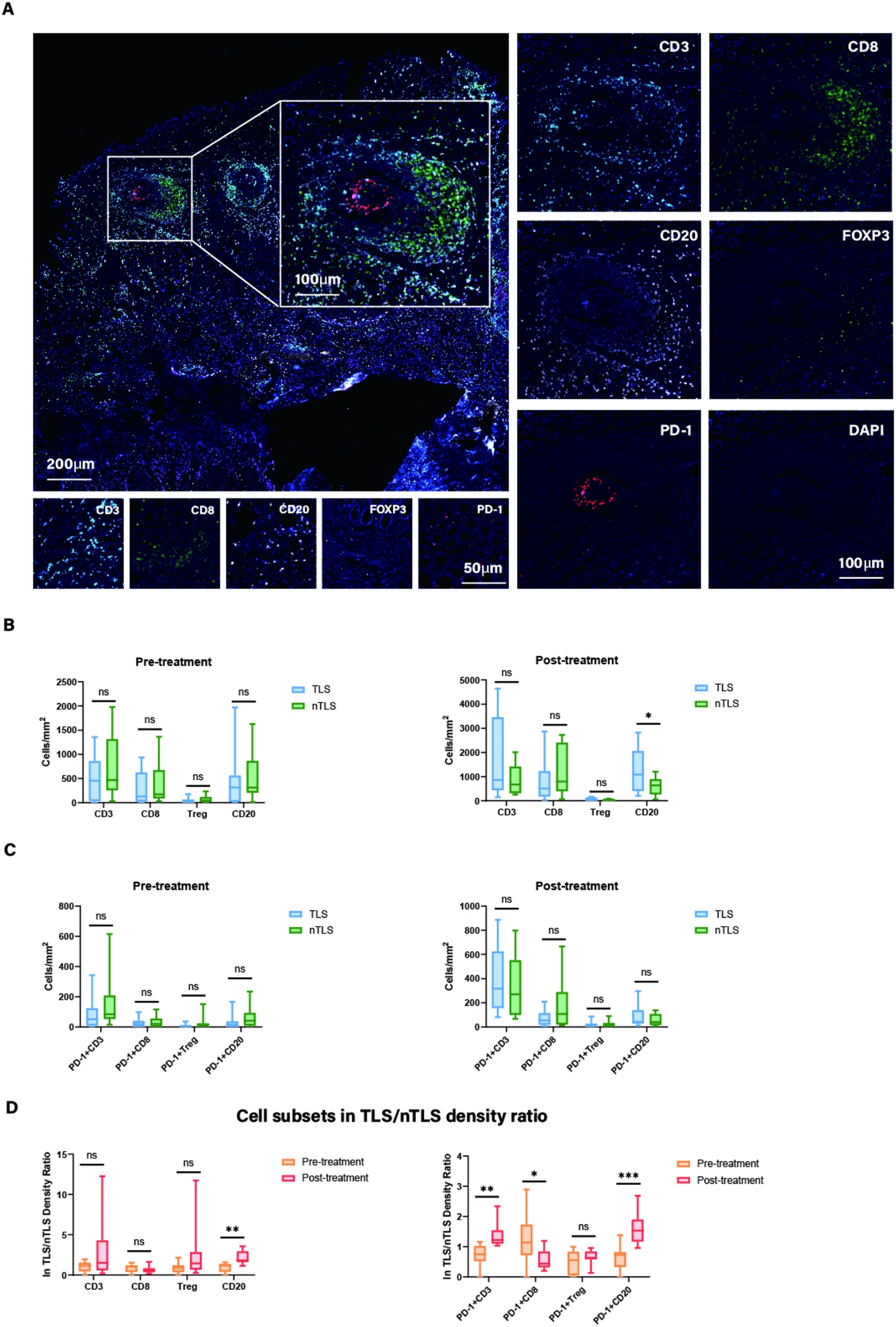
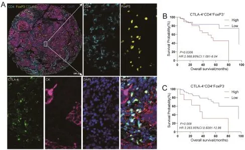
The Role of PD-1+ Treg/PD-1+ CTL Ratio and Tertiary Lymphoid Structures in Gastric Cancer
A new study shows PD-1+ Treg/CTL ratio combined with TLS predicts prognosis and treatment response in gastric cancer using mIF, TissueFAXS, and StrataQuest.
Customer Publication
15 Apr, 2025
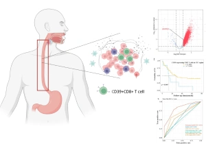
CD39+CD8+ T Cells as a Marker for Diagnosis and Prognosis of ESCC
A research group in China analyzed CD39+CD8+ T cells in esophageal squamous cell carcinoma. Based on mIF-stained samples acquired on the TissueFAXS platform, they propose these cells as new markers for diagnosis and prognosis.
Application Note
11 Sep, 2024
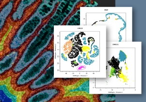
A Glimpse into StrataQuest 8: Spatial Analysis in Colorectal Cancer
The newest StrataQuest supports in-depth neighborhood analysis with exportable data for statistics. It includes manifold learning (t-SNE, UMAP, SONG), violin plots, 3D diagrams, and other tools that give full control of data.
Webinar
20 Aug, 2024
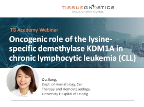
Oncogenic Role of KDM1A in Chronic Lymphocytic Leukemia (CLL)
Dr. Qu Jiang (Leipzig) presents new findings on the oncogenic role of KDM1A in CLL. Using StrataQuest, her team quantified IF-stained murine spleen sections to assess leukemic burden after Kdm1a knockdown.
Customer Publication
06 Aug, 2024
Unlocking the Potential of HHLA2 in Laryngeal Squamous Cell Carcinoma
Researchers investigated HHLA2 as a prognostic marker in LSCC using multiplexed IHC on TMA cores. TissueFAXS automated imaging and StrataQuest analysis revealed HHLA2 expression across immune cell populations.
Webinar
10 May, 2024
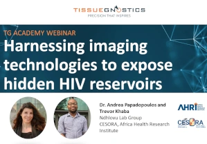
Harnessing Imaging Technologies to Expose Hidden HIV Reservoirs
Dr. Andrea Papadopoulos and Trevor Khaba from Ndhlovu group, CESORA, Africa Health Research Institute, present their latest work on finding and characterizing new HIV reservoirs.

Application Note
15 Jan, 2024
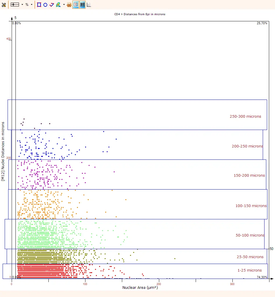
Spatial Analysis of CD4+ Cells in Colorectal Cancer
StrataQuest contextual image analysis reveals how CD4+ T helper cells interact with their native tumor microenvironment in colorectal cancer, a leading malignancy in Europe.
White Paper
25 Oct, 2023
Characterizing the Tumor Microenvironment of Colorectal Cancer Patients
A rare MSS-type CRC patient responded to immunotherapy. TissueFAXS SL and StrataQuest were used to image and quantify immune phenotypes.
Customer Publication
13 Jul, 2023
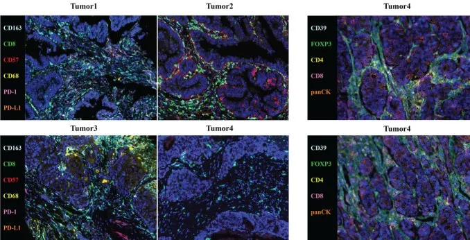
Tumor Microenvironment Characterization of Rectal Cancer
Song et al. investigated a rectal cancer case with MSS/PD-L1-negative recurrent hepatopulmonary metastasis. Using TissueFAXS and StrataQuest, they characterized tumor-infiltrating immune cells, including CD4, CD8, CD163, PD1, and PD-L1.
Customer Publication
08 Nov, 2022
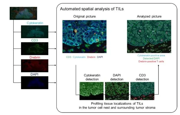
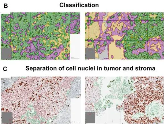
Characterization of the Spatial Immune Context of Lung Cancer
Imamura et al. characterized debrin+ tumor-infiltrating lymphocytes in lung cancer using StrataQuest for automated classification and spatial localization of immune phenotypes in tumor and stroma.
Customer Publication
08 Nov, 2022
Expression and clinical significance of VISTA, B7-H3, and PD-L1 in glioma
Wang et al. investigated RNA and protein expression of immune checkpoint molecules in glioma, highlighting prognostic value. TissueFAXS SPECTRA enabled multispectral TMA scanning, and StrataQuest was used for in-depth phenotyping.
White Paper
12 Oct, 2022
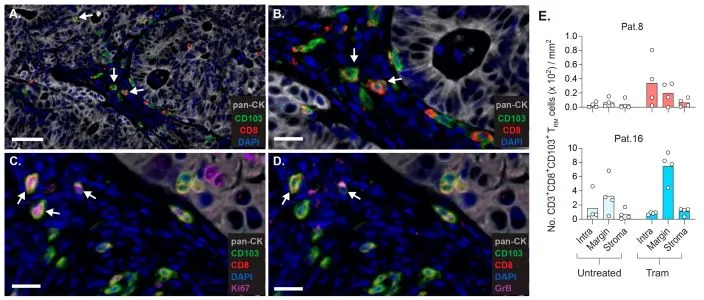
CD8+ TRM Cells as Potential Targets in Colorectal Liver Metastases
A study using TissueFAXS CHROMA and StrataQuest revealed that CD103+CD39+CD8+ TRM cells accumulate in colorectal liver metastases compared to adjacent liver tissue. These findings highlight their potential as therapeutic targets in advanced colorectal cancer.
Customer Publication
05 Oct, 2022
EMT-Transcription Factors as Drivers for Recurrence in HNSCC
Ingruber et al. examined pEMT-related proteins driving cisplatin resistance and recurrence in HNSCC. TissueFAXS and StrataQuest were applied for quantitative tissue cytometry and phenotyping.
Customer Publication
14 Sep, 2022
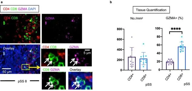
Cytotoxic CD8+ T cell Role in Tissue Destruction in Sjögren’s Syndrome
Kaneko et al. (Scientific Reports, Nature) investigated CD4+ and CD8+ T-cell subsets in salivary glands of Sjögren’s syndrome patients using TissueFAXS with TissueQuest and StrataQuest for in-depth quantitative tissue cytometry.
Blog Post
01 Sep, 2022
What is Spatial Phenotyping?
Spatial phenotyping combines whole-slide imaging and advanced analysis to study cellular phenotypes in their native context. This approach is key in immunotherapy research, enabling insights into the tumor microenvironment and supporting informed treatment strategies.