IF Pyknotic Nuclei
Detect tissue, segment cells, and identify pyknotic nuclei by nuclear staining, define IF-stained phenotypes, perform dot detection, segment nucleus/perinuclear area/cytoplasm, and export counts, mean intensity etc.
fluorescence
single-cell analysis
cell culture
pyknotic nuclei, chromatin, necrosis, apoptosis, cell culture, fluorescence
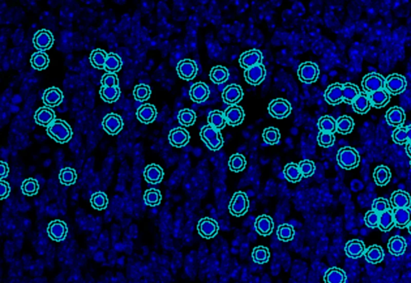
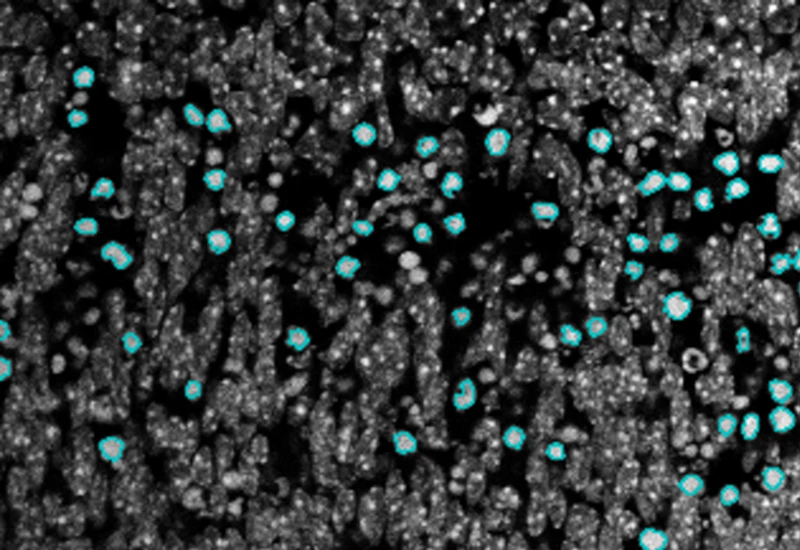
The IF Pyknotic Nuclei App provides tissue detection and cell segmentation in combination with the detection of pyknotic nuclei (defined as completely condensed, round, high intensity nuclei) based on nuclei staining. Additionally, the APP allows to determine the cellular phenotype of specific IF stained cell populations as well as dot detection. It segments the detected cells into nucleus, and/or perinuclear area and/or cytoplasm. As outcome the APP provides parameters such as number, mean intensity and percentage of specific cell populations (including cells containing pyknotic nuclei), as well as dot parameters per segmented cell including count, mean intensity, total dot area, and sum of intensity as well as area and intensity list for all single dots.
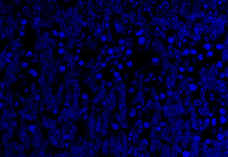
Original image
Pyknotic nuclei detection
cell culture

Webinar
19 Nov, 2024
Quantification of p-H2AX Foci in Co-cultured Cells Exposed to Radiation, Livia Sima
single-cell analysis
White Paper
30 Mar, 2026
Understanding NeuroCOVID-19: SARS-CoV-2 Disrupts Astrocyte Homeostatic Functions
fluorescence
Blog Post
07 Apr, 2026
How immunofluorescence image analysis factors into NSCLC studies
We support the following file formats:
- TissueFAXS (aqproj)
- StrataFAXS II (vmic)
- PreciPoint (vmic, gtif)
- Generic BigTIFF Import
- Support for multipage BigTIFF files
- OME-TIFF
- JPEG, PNG, BMP, TIFF
- Zeiss (czi)
- Hamamatsu NanoZoomer (ndpi)
- Aperio (svs)
- Leica (scn)
- 3D HISTECH Pannoramic
- Mirax (mrxs)
- Olympus (vsi)
- More slide scanners to be added!
Related Apps

IF Cardio Cell Culture Dots
Segment cultured cardiac cells, detect cardiomyocytes and fibroblasts, and quantify dot markers (CISH/FISH) per cell, including cell counts and dot number, area, and mean intensity.
cardiology, cardiomyocytes, cell culture, fibroblasts, troponin red, FISH

IF Cultured Cells & Substructures
Detect nuclei-based cells, quantify dot markers per cell, and measure cytoskeletal filament density and dot intensity at single-cell level.
FISH, cell culture, cytoskeleton, actin fibres
.webp)
IF Leishmaniasis
Detect intracellular Leishmania in host cells, segment parasites, quantify parasites per cell, distinguish live/dead (specifc assay), and export 20 intensity, statistic, and morphometric parameters per compartment plus parasite count, mean intensityand size.
macrophages, parasites, leishmaniasis, cell culture, protozoan parasites, fluorescence

Custom App development
Perfectly tailored image analysis solutions for your research.
You have a specific research question that needs to be answered? We offer custom development of image analysis pipelines for specific tasks, be it detection of cellular phenotypes or quantification of tissue structures. After discussing your goals with one of our experts, you will get a ready-to-use App and be a step closer to an impactful publication.