IF Neurite
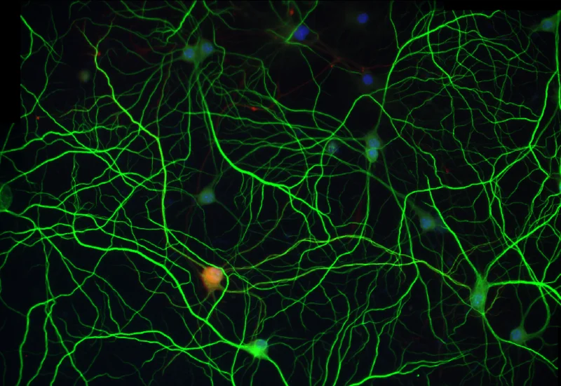
Detect neuronal cells and clusters with their neurites/dendrites, quantify branching per neuron, and export total area, length, average thickness, branch points, and endpoints.
single-cell analysis
neuroscience
cell culture
cell culture, fluorescence, neurites
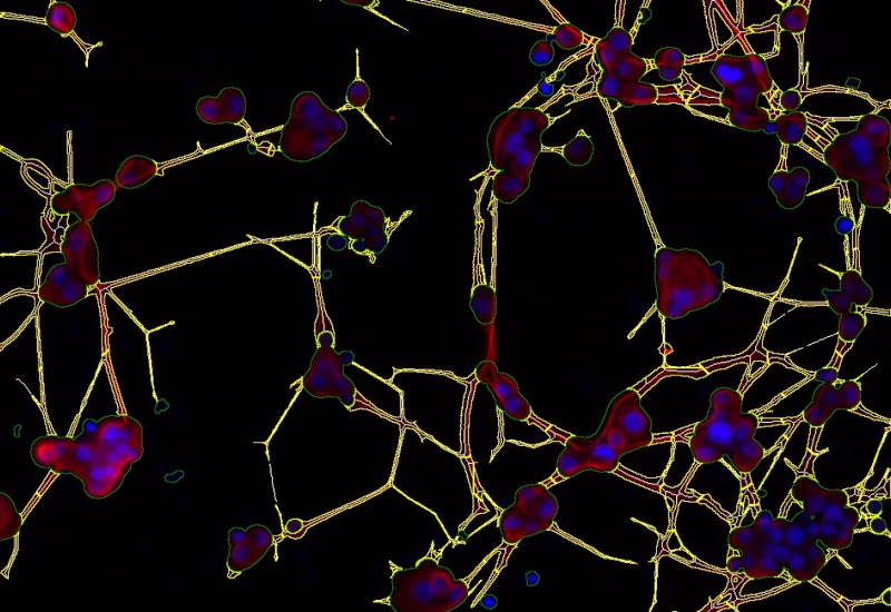
The IF Neurite App identifies neuronal cells and cell clusters and their neurites/dendrites. It quantifies the number of neurites/dentrites branching out from a specific neuron, identifies branch points and exports total neurite/dentrites area, total neurite/dentrites length, average neurite/dentrites thickness, number of branch points, and number of end points.

Original image
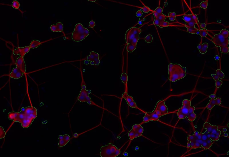
Cell body detection
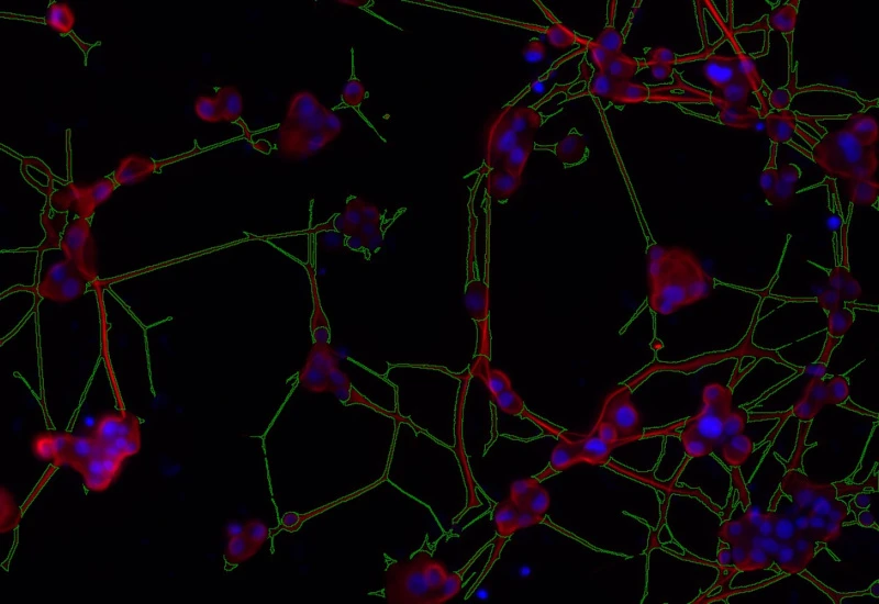
Neurite/dendrite detection
Nuclei/neurite/dendrite detection
cell culture

Webinar
19 Nov, 2024
Quantification of p-H2AX Foci in Co-cultured Cells Exposed to Radiation, Livia Sima
neuroscience
White Paper
30 Mar, 2026
Understanding NeuroCOVID-19: SARS-CoV-2 Disrupts Astrocyte Homeostatic Functions
single-cell analysis

Blog Post
23 Jul, 2026
How Histology Slide Scanners are Used to Study Osteoarthritis
We support the following file formats:
- TissueFAXS (aqproj)
- StrataFAXS II (vmic)
- PreciPoint (vmic, gtif)
- Generic BigTIFF Import
- Support for multipage BigTIFF files
- OME-TIFF
- JPEG, PNG, BMP, TIFF
- Zeiss (czi)
- Hamamatsu NanoZoomer (ndpi)
- Aperio (svs)
- Leica (scn)
- 3D HISTECH Pannoramic
- Mirax (mrxs)
- Olympus (vsi)
- More slide scanners to be added!
Related Apps
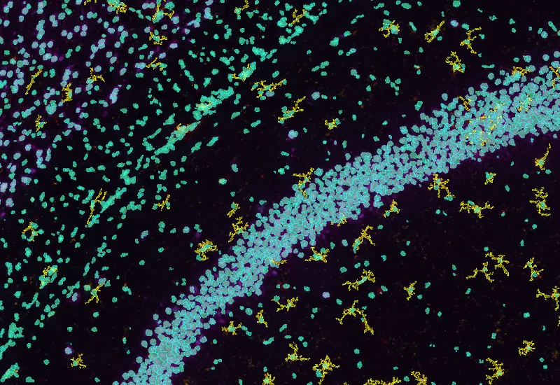
IF Brain App
Classify brain regions using an AI-based classifier and detect cellular phenotypes (e.g., astrocytes) based on markers, quantify tissue areas, total cells, and phenotype counts across regions.
brain, mouse, astrocytes, machine learning
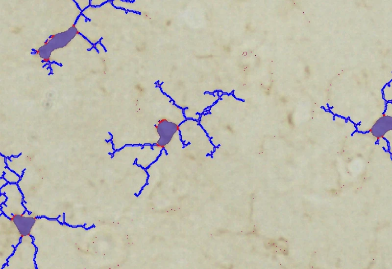
IHC Microglia
Detect microglia soma in IHC-stained samples, segment branches, and identify primary and secondary branching points to quantify cell morphology and branching complexity.
microglia, central nervous system, peripheral nervous system, phagocytosis, astrocytes, branches

Custom App development
Perfectly tailored image analysis solutions for your research.
You have a specific research question that needs to be answered? We offer custom development of image analysis pipelines for specific tasks, be it detection of cellular phenotypes or quantification of tissue structures. After discussing your goals with one of our experts, you will get a ready-to-use App and be a step closer to an impactful publication.