Latest articles in

White Paper
01 May, 2025
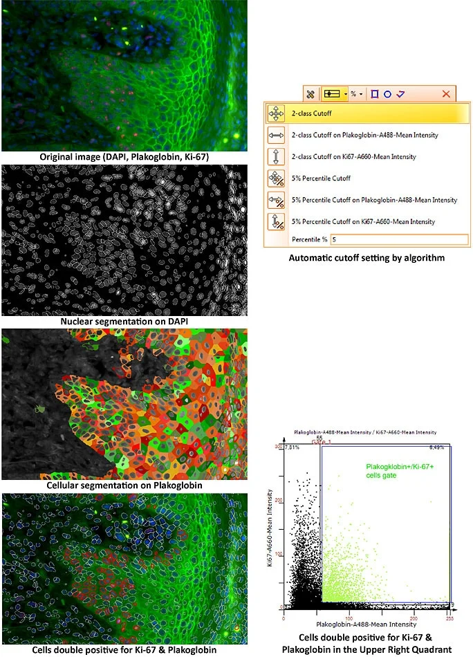
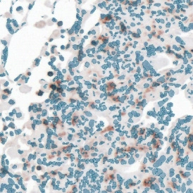
Epigenetic Carcinogenesis: Quantitative Insights from HistoQuest Analysis
The evolving understanding of cancer biology underscores the pivotal role of epigenetic modifications - heritable yet reversible changes in gene expression that do not alter the DNA sequence.
Webinar
05 Feb, 2024
Unveiling the Dichotomous Role of STAT3 Signaling in Prostate Cancer
Dr. Lukas Kenner presents his research on STAT3’s dual role in prostate cancer. TissueFAXS, HistoQuest, and StrataQuest enabled whole-slide imaging and single-cell quantification.
News
16 Oct, 2023
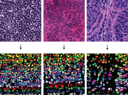
Deep Learning for Improved Nuclei Segmentation in Microscopic Images
Dr. Amirreza Mahbod presents how deep learning enhances nuclei segmentation and tackles challenges in histological image analysis.
Blog Post
09 Oct, 2023
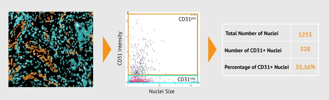
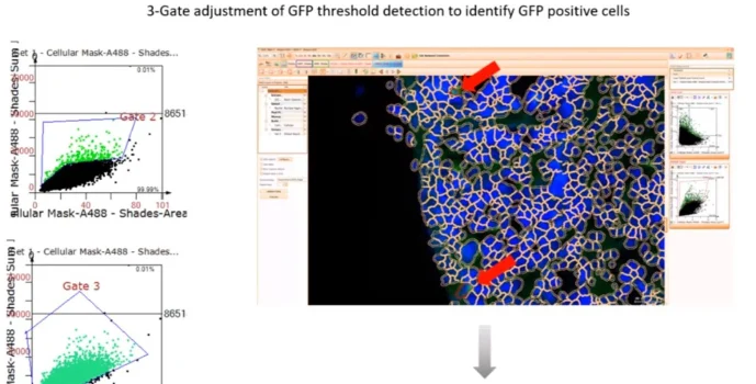
Revolutionizing Tissue Analysis: Image-Based Cytometry
Image-based cytometry is transforming tissue analysis by combining microscopy with flow cytometry features. It enables rapid, automated characterization of cells in their native environment, advancing pathology, oncology, and precision medicine with AI integration.
White Paper
12 Jun, 2023
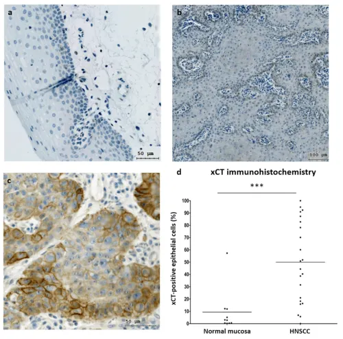
Activated Erk 1/2 Kinases Decrease Cell Viability Caused by Erastin in HNSCC
A recent study using TissueFAXS and HistoQuest revealed that xCT inhibition by erastin may trigger ferroptosis in HNSCC cells. Erk1/2 activation sensitized tumor cells to this process, suggesting novel therapeutic strategies for head and neck cancer.
Blog Post
23 Jan, 2023
An Introduction to Spatial Tissue Cytometry
Spatial tissue cytometry preserves cells in their native environment, enabling single-cell analysis and spatial insights into tissue organization. Discover how TG’s imaging and analysis platforms unlock powerful applications in research and precision medicine.
Webinar
12 Oct, 2022
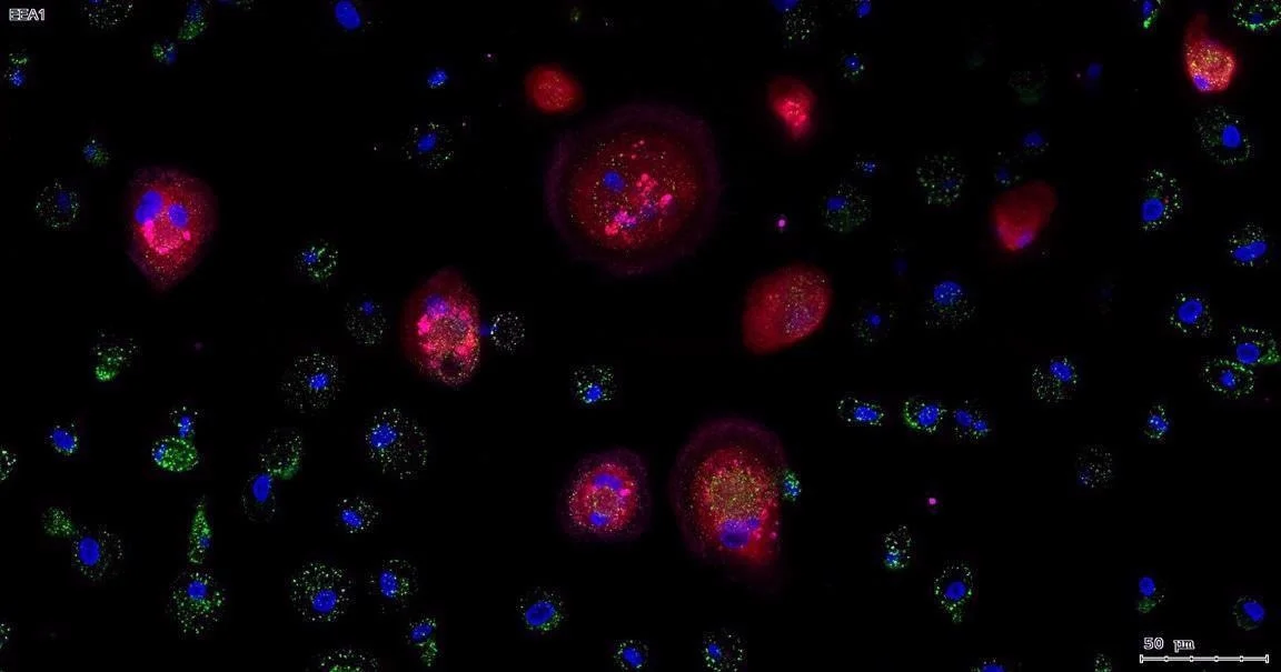
Quantitative Image-based Cytometry: Focus on the Foci!
Prof. Nicolas C. Hoch, University of Sao Paulo, is presenting his research on signaling and repair of DNA damage. He and his group are utilizing the power of TissueFAXS i PLUS slide scanning and the high-end image analysis solution, StrataQuest, for their research.
Application Note
04 Oct, 2022
High-content phenotyping with Imaging Mass Cytometry
StrataQuest enables high-content image analysis of IMC datasets. This case study demonstrates single-cell and proximity measurements across 30 markers within one tissue section for comprehensive phenotyping.
White Paper
15 Jun, 2022
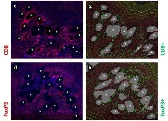
Comprehensive Tissue Cytometry Analysis of CD3+ T cells in Lungs of COVID-19 Patients
This white paper details the first in-depth tissue cytometry analysis of CD3+ T-cell subsets in COVID-19 lungs. Using TissueFAXS PLUS and StrataQuest, researchers uncovered dysfunctional CD8+ T cells and expansion of cytotoxic CD4+ T cells driving impaired immunity.
Interview
11 May, 2022
Disrupted B and T Cell Dynamics in COVID-19 Lymph Nodes Revealed by Tissue Cytometry
Dr. Thomas Diefenbach and colleagues at the Ragon Institute analyzed B and T cell populations in COVID-19 patient lymph nodes using TissueFAXS and StrataQuest. Their microscopy and image analysis revealed disrupted germinal centers and impaired humoral immunity.
Webinar
06 Apr, 2022
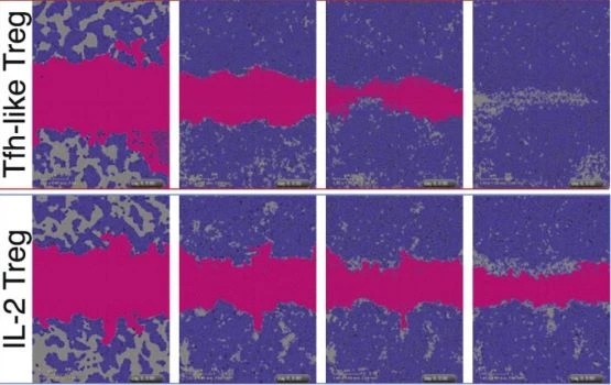
Analysis of tumour initiating cells and syngeneic EPSC-derived neural stem cells in glioblastoma
Claire Vinel, PhD (Queen Mary University London), presents her Nature Communications study on epigenetic mechanisms in glioblastoma. The work highlights Treg recruitment via glycosaminoglycans and identifies druggable, patient-specific targets using integrated transcriptome and methylome analysis.
Customer Publication
08 Feb, 2022
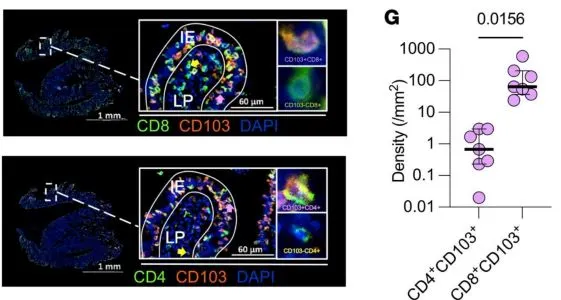
Differential localization and limited cytotoxic potential of duodenal CD8+ T cells
A study in The Journal of Clinical Investigation (Mvaja et al.) shows the limited potential of duodenal CD8+ T cells in people living with HIV. High-content immunophenotyping was performed using TissueFAXS i PLUS and TissueQuest single-cell analysis.

Blog Post
07 Dec, 2021
What is Multispectral Cytometry?
Multispectral cytometry resolves spectral overlap to enable accurate, high-parameter analysis of cells and tissues. Learn how this technology, paired with spectral unmixing and advanced imaging, expands applications from cancer research to immunotherapy.
Customer Publication
12 Oct, 2021
Tissue Cytometry in HIV Research
Collins et al. (Ragon Institute, Harvard/MIT/MGH) reveal key virologic and immunologic factors of durable HIV control. HIV+ lymph nodes were visualized via RNAScope and acquired using TissueFAXS Q (Cell Press).