
Case Study: Analysis of FISH Using Tissue Cytometry
The IF Dots App in StrataQuest streamlines automated analysis of FISH stainings by detecting nuclei and quantifying dots per cell. This case study shows how the workflow enables accurate, exportable data for genetics research and clinical applications.
image analysis
FISH analysis
digital pathology
Application Note
Case Study: Analysis of FISH Using Tissue Cytometry
14 Feb, 2023
Fluorescence in situ hybridization (FISH) has been routinely used for decades in research and clinics to rapidly identify potential genetical changes within cells. This case study shows how TissueGnostics’ image analysis solution StrataQuest can be applied to evaluate FISH stainings. The most streamlined solution, the IF Dots App (runs within StrataQuest), can be applied to any analysis of dot-sized objects within the nuclei. Depending on the research needs, the following analysis can be combined with any other kind of analysis including AI-based tissue classification, phenotyping, spatial analysis, etc.
The aim of the following case study was to (i) identify individual nuclei and (ii) to assess how many FISH dots per individual nuclei are present.
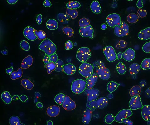
The image below shows an overview of a triple staining: the nucleus stained by DAPI (blue), and two FISH markers, the housekeeping gene (green), and HER2/neu (red).

The automated analysis starts with nuclei segmentation (a), where nuclei are identified by the intensity of the DAPI staining (nuclei are marked by the green outline). In the next step, the FISH dots are detected: in red the housekeeping gene in FITC channel (b) and in yellow for the HER2/neu in Acridine channel (c). Then these masks can be combined, as seen in (d).

For further visualization of the analysis results, StrataQuest outputs the data in scattergrams. In the example below, the number of Acridine dots per nucleus (y-axis) is plotted against the FITC dots per nucleus (x-axis). The identified nuclei were gated for the condition “2 acridine dots per nuclei”, where the number of FITC dots can vary, and selected cells are shown in red contour via backward connection. Scattergrams showing the number of detected nuclei with each dot number combination are useful, especially with the Heat Map function enabled (bottom) which indicates those groups with a higher quantity of nuclei. To gain quantitative data for statistical analysis, the results can be immediately exported to Excel, CSV, and PDF formats.

FISH analysis is only one example of StrataQuest´s capabilities. If you are interested in how StrataQuest streamlined image analysis solution can enhance your research today, please reach out to a TissueGnostics team member for a demonstration.
Get to know more about FISH applications combined with TissueGnostics solutions in our blog posts:
Latest Articles
Blog Post
11 Nov, 2026
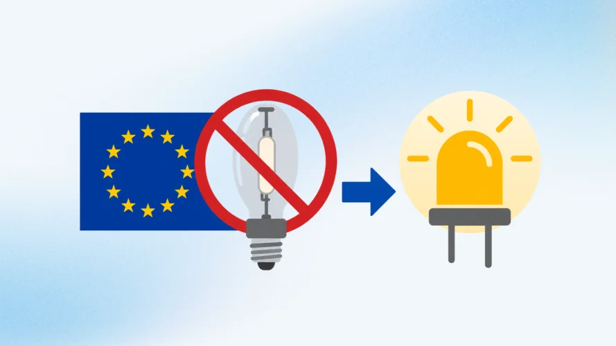
EU rules on mercury and metal-halide lamps: what microscopy labs need to know
EU regulations on mercury-containing lamps are changing, raising questions about replacement bulbs and long-term microscope maintenance. Find out what the February 2027 exemption deadline means, how it affects existing microscopy systems, and why now is a good time to consider switching to LED illumination.
White Paper
01 Oct, 2026
A New Neutrophil Subset Suppresses Immunity Response in CRCLM
Within the tumor microenvironment, tumor-associated neutrophils (TANs) are gaining attention as important regulators of inflammation, immune suppression, and tissue remodeling. Recent studies suggest that specific neutrophil populations can actively support metastatic growth rather than simply responding to it.
News
14 Sept, 2026
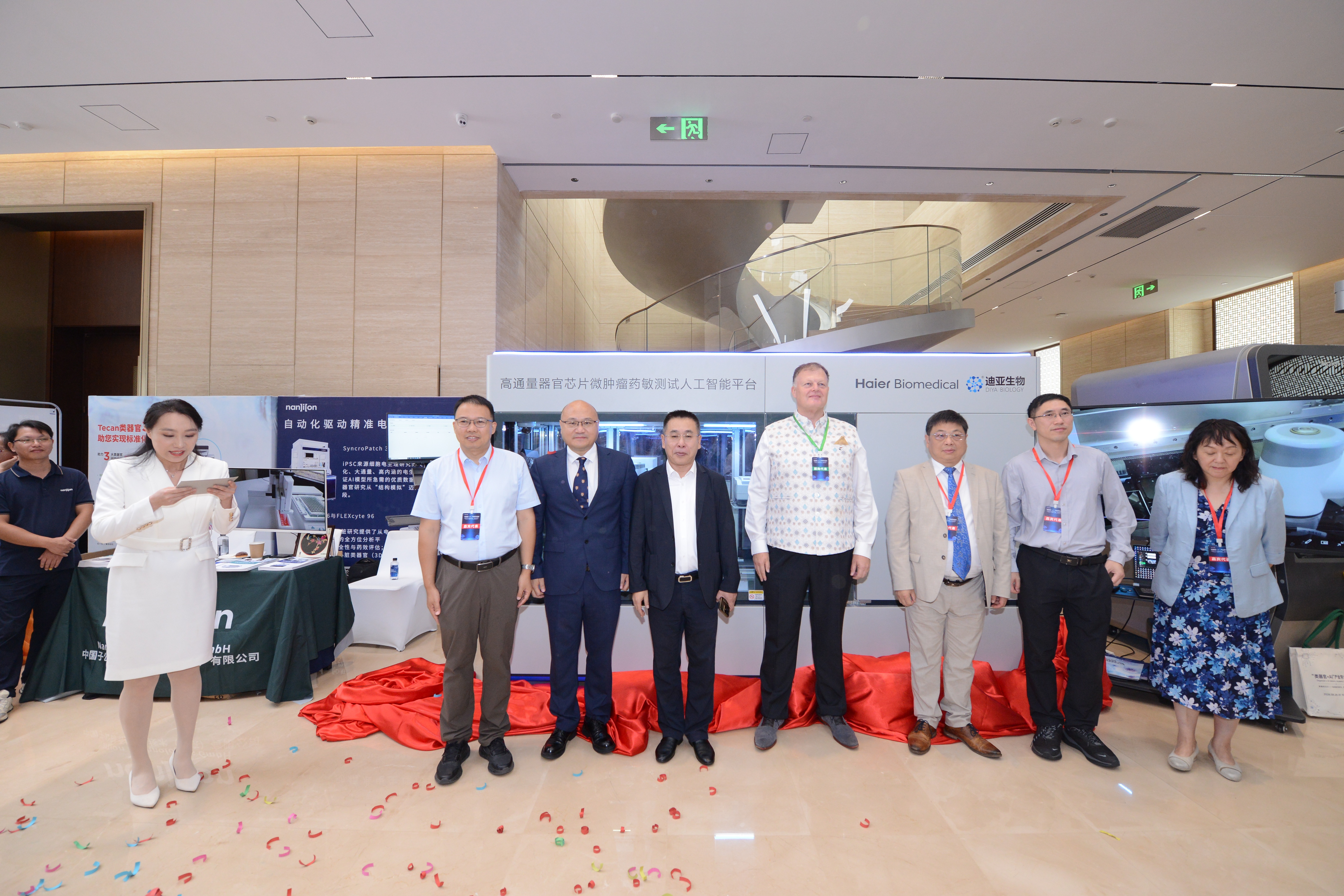
TissueGnostics collaborates with Diya Biological Technology in organoid and organ-on-a-chip innovation
TissueGnostics, the trailblazer in Spatial Tissue Cytometry, has entered into a collaborative agreement with Diya Biological Technology Group Co., Ltd., a leading Chinese organ-on-a-chip CDMO and research service platform specialising in microfluidic organ chips, organoid banking and tumour drug-testing services.

