
A New Neutrophil Subset Suppresses Immunity Response in CRCLM
Within the tumor microenvironment, tumor-associated neutrophils (TANs) are gaining attention as important regulators of inflammation, immune suppression, and tissue remodeling. Recent studies suggest that specific neutrophil populations can actively support metastatic growth rather than simply responding to it.
spatial analysis
tumor microenvironment
multiplex IF
immunology
White Paper
A New Neutrophil Subset Suppresses Immunity Response in CRCLM
01 Oct, 2026
Colorectal cancer with liver metastasis (CRCLM) is the leading cause of CRC-associated death. Although surgery, chemotherapy, targeted therapies, and immunotherapy have improved outcomes, many patients with colorectal cancer liver metastasis still experience progressive disease.
Increasing evidence indicates that the tumor microenvironment plays a central role in controlling therapeutic response, immune activity and metastatic progression. Within this complex ecosystem, tumor-associated neutrophils (TANs) are gaining attention as important regulators of inflammation, immune suppression, and tissue remodeling. Recent studies suggest that specific neutrophil populations can actively support metastatic growth rather than simply responding to it (1,2).
Revealing Cellular Diversity through Single Cell Analysis
Traditional methods often classify neutrophils as a single immune population, yet advanced single cell technologies such as scRNA-seq have revealed substantial diversity within these cells. Distinct neutrophil subsets can display different transcriptional programs, functional properties, and interactions with neighboring cells. In CRCLM, researchers identified a neutrophil population characterized by expression of TAN1, LGALS3 and genes associated with hypoxia, angiogenesis and immune regulation. This discovery highlights how metastatic tissues can shape immune cell behavior. Understanding these specialized cellular states is important because individual neutrophil subsets may influence tumor progression, immune escape and response to therapy in different ways (1,3).
Spatial Biology Adds Essential Context
While single-cell RNA sequencing provides valuable information about cellular identity, it does not fully explain how cells are organized within tissues. Spatial biology approaches address this limitation by linking molecular information to precise tissue locations.
In metastatic liver lesions, researchers observed that distinct neutrophil populations accumulate within specific regions of the tumor microenvironment. These spatial patterns suggest that local conditions such as oxygen availability, vascular architecture, and interactions with neighboring immune cells influence neutrophil function. Mapping these relationships provides deeper insight into how metastatic niches develop and why certain microenvironments become particularly supportive of tumor growth and persistence (1,4).
Multiplex Immunofluorescence Confirms Biological Findings
While scRNAseq delivers important insights into key genomic differences between cells, multiplex immunofluorescence can not only validate these observations, but add a spatial layer of additional information.
Multiplex immunofluorescence enables simultaneous visualization of multiple biomarkers within a single tissue section while preserving tissue architecture. This capability allows researchers to directly observe cellular relationships that cannot be captured using conventional single-marker techniques.
By confirming the localization and phenotype of critical immune populations, multiplex imaging provides an important bridge between molecular data and the actual biology. Such validation is essential when developing a comprehensive understanding of the tumor microenvironment and its influence on metastatic disease progression (1,5).
A Unique Neutrophil Phenotype Emerges
Multiplex imaging confirmed that TAN1_LGALS3 neutrophils possess a distinctive phenotype characterized by high CXCR4 expression and comparatively low CXCR1 and CXCR2 levels (1). Importantly, researchers observed significantly greater infiltration of CXCR4-positive neutrophils in liver metastases than in primary colorectal tumors, supporting the conclusion that this population performs specialized functions within metastatic niches.
Additional analyses revealed expression patterns associated with hypoxic adaptation, metabolic reprogramming, and pro-angiogenic activity, suggesting that these cells help establish conditions favorable for tumor growth persistence, vascular development, and resistance to immune-mediated destruction within advanced metastatic disease settings affecting patient outcomes, clinical management, and future therapies.
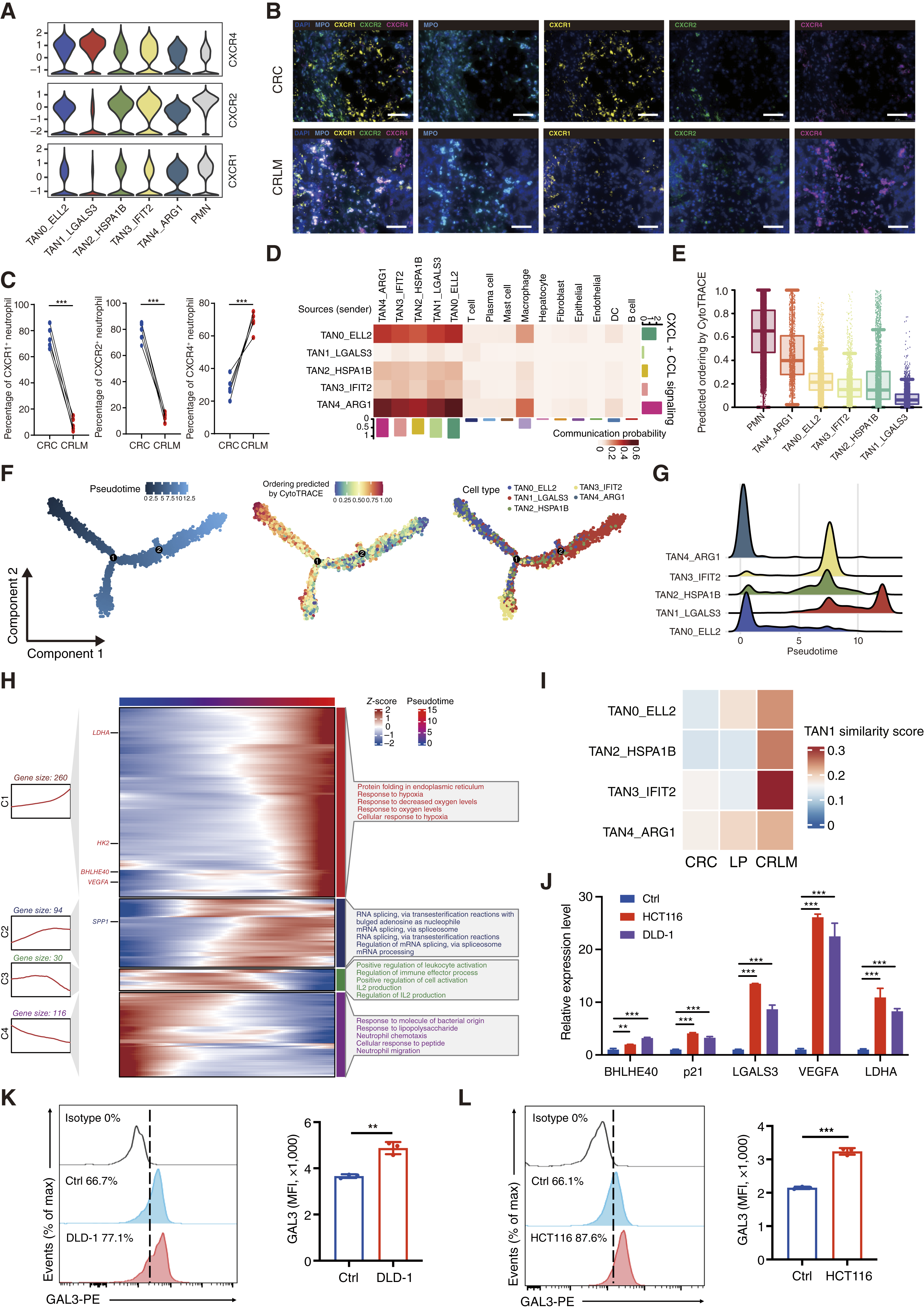
From Figure 3. Tissue specification of neutrophils in the CRLM microenvironment. Zhihang, C., et al. (2026). Senescent-like neutrophils shape angiogenic immunosuppressive niches in colorectal cancer liver metastasis. Cancer Discov, 10.1158/2159-8290.CD-25-0820
Tyramide Signal Amplification for Better Signal
The detection of low-abundance targets can be challenging in complex tumor tissues. To overcome this limitation, researchers employed Tyramide Signal Amplification (TSA) Multiplex immunohistochemistry (IHC) technology from TissueGnostics.
TSA chemistry amplifies fluorescent signals while maintaining high spatial resolution and marker specificity. This approach enables reliable detection of weakly expressed biomarkers and supports the construction of highly multiplexed staining panels. For studies investigating numerous cellular populations simultaneously, signal amplification is particularly valuable because it improves sensitivity without sacrificing image quality. As a result, investigators can generate more complete datasets and gain a clearer understanding of cellular interactions occurring within metastatic lesions.
High-Quality Imaging without Spectral Interference
Accurate multiplex analysis depends on high-quality image acquisition. TissueFAXS Spectra, an automated slide scanner, was used to capture publication-ready images from stained tissue sections.
Designed specifically for multiplex fluorescence (mIF) applications, the platform incorporates spectral unmixing capabilities that help eliminate autofluorescence and channel bleed-through.
These features are particularly important when analyzing tissues containing multiple overlapping fluorescent signals. With support for multiple filter sets and automated scanning workflows, TissueFAXS Spectra enables researchers to produce consistent high-resolution images while maximizing throughput. Reliable image acquisition forms the foundation for meaningful analysis and confident biological interpretation.
Quantitative Analysis with StrataQuest
Obtaining images is only part of the scientific workflow. Researchers must also extract meaningful quantitative information from the resulting datasets.
In this study, StrataQuest software was used to perform image processing and advanced spatial analysis (1). The platform supports cell segmentation, phenotyping, density measurements, and evaluation of cell-to-cell relationships across entire tissue sections.
By transforming complex images into quantitative data, StrataQuest enables researchers to compare biological features objectively and statistically. This capability was critical for validating observations generated by single-cell and spatial transcriptomic analyses and for identifying functionally important cellular interactions within metastatic tissues.
Formation of Angiogenic Immunosuppressive Niches
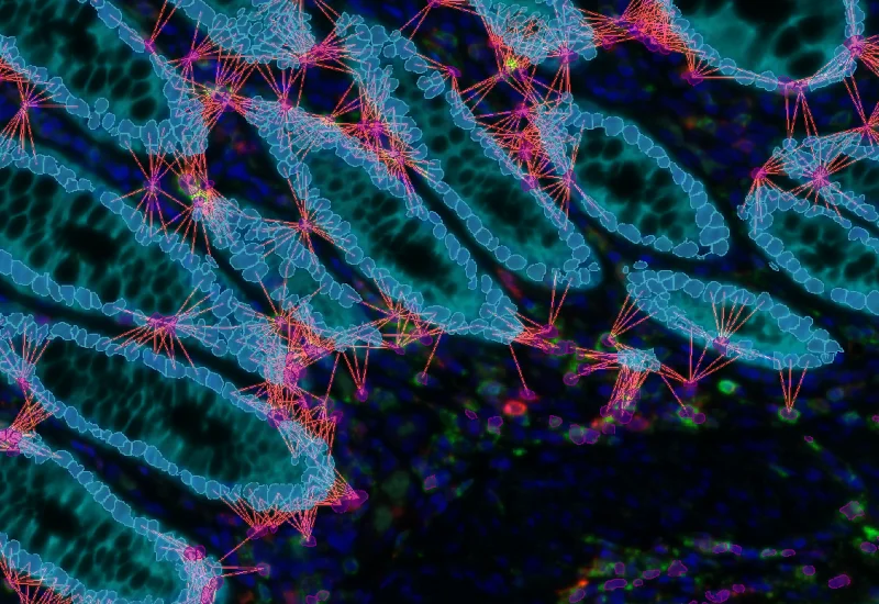
One of the most important discoveries was the identification of organised cellular niches composed of multiple interacting cell types. Within colorectal cancer liver metastases, researchers observed the colocalization of BHLHE40+p21+neutrophils, CD68+ macrophages, and CD31+ vascular endothelial cells (1).
These structures were rarely detected in primary colorectal tumors but appeared frequently in metastatic lesions. Their presence suggests coordinated biological activity involving angiogenesis, immune suppression, and tumor support mechanisms. By existing in close proximity, these cellular partners may exchange signals that collectively strengthen the metastatic microenvironment and promote progression despite therapeutic intervention and ongoing host immune responses (1,6).
Linking Transcriptomics, Imaging and Spatial Biology
One of the most significant aspects of this work is the integration of multiple complementary technologies. Single-cell sequencing identified biologically relevant neutrophil populations. Spatial transcriptomics revealed their location within the tissue microenvironment. Multiplex immunofluorescence confirmed these observations in situ on the protein level, while advanced image analysis provided quantitative support.
The combination of these methodologies generated a multidimensional view of metastatic biology that would not have been possible using any single technology alone. This integrated approach demonstrates the value of connecting molecular discoveries to visual and spatial evidence when investigating complex diseases such as colorectal cancer metastasis.
Future Direction
Collectively, the data indicate that senescent-like TAN1_LGALS3 neutrophils are critical in influencing angiogenic, immune-suppressive microenvironments that facilitate colorectal cancer liver metastasis. By combining single-cell technologies with advanced multiplex immunofluorescence, researchers uncovered critical cellular interactions that may influence disease progression and therapeutic response. The findings demonstrate how sensitive staining methods, high-quality imaging, and sophisticated image analysis can reveal complex biological relationships within intact tissues, opening new opportunities for future biomarker discovery and therapeutic development.
References and Further Reading
1. Zhihang, C., et al. (2026). Senescent-like neutrophils shape angiogenic immunosuppressive niches in colorectal cancer liver metastasis. Cancer Discov, 10.1158/2159-8290.CD-25-0820
2. Galdiero, M. R., et al. (2021). Occurrence and significance of tumor-associated neutrophils in patients with colorectal cancer. International Journal of Cancer, 149(2), 446–456. 10.1002/ijc.30076
3. Jaillon, S., et al. (2020). Neutrophil diversity and plasticity in tumor progression and therapy. Nature Reviews Cancer, 20(9), 485–503. 10.1038/s41568-020-0281-y
4. Binnewies, M., et al. (2018). Understanding the tumor immune microenvironment and its therapeutic implications. Nature Medicine, 24(5), 541–550. 10.1038/s41591-018-0014-x
5. Stack, E. C., et al. (2014). Multiplexed immunohistochemistry, imaging, and quantitation: A review, with an assessment of tyramide signal amplification, multispectral imaging and multiplex analysis. Methods, 70(1), 46–58. 10.1016/j.ymeth.2014.08.016
6. Carmeliet, P., & Jain, R. K. (2011). Principles and mechanisms of vessel normalization for cancer and other angiogenic diseases. Nature Reviews Drug Discovery, 10(6), 417–427. 10.1038/nrd3455

