Latest articles in
White Paper
01 Jun, 2024
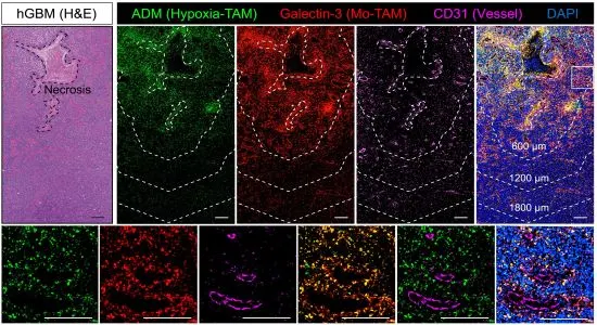
Understanding Glioblastoma, One Cell at a Time
Wang et al. used spatial transcriptomics to characterize subsets of tumor-associated macrophages (TAMs) known as monocyte-derived TAMs. They identified a cluster of hypoxia-TAM oversecreting adrenomedullin, confirmed by multiplex immunostaining with TissueFAXS Spectra imaging platform, and StrataQuest image analysis software was employed for marker identification.

Webinar
10 May, 2024
Harnessing Imaging Technologies to Expose Hidden HIV Reservoirs
Dr. Andrea Papadopoulos and Trevor Khaba from Ndhlovu group, CESORA, Africa Health Research Institute, present their latest work on finding and characterizing new HIV reservoirs.
White Paper
04 Apr, 2023
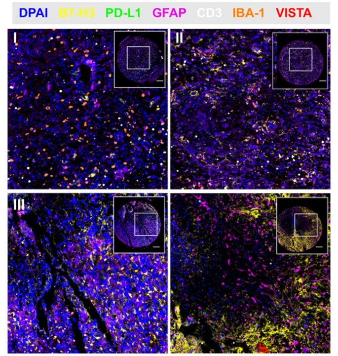
In-Depth Immune Checkpoint Analysis of Glioma at Transcript and Protein Level
Researchers from Sichuan University performed transcriptomic and multiplex immunofluorescence analyses of glioma samples using TissueFAXS SPECTRA and StrataQuest. Findings highlight B7-H3 as a prognostic biomarker and potential immunotherapy target in glioma.
Customer Publication
08 Nov, 2022
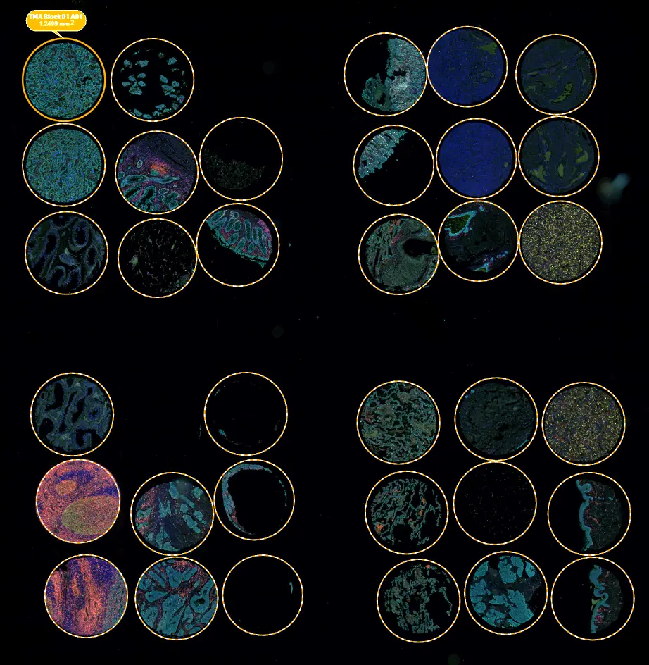
Expression and clinical significance of VISTA, B7-H3, and PD-L1 in glioma
Wang et al. investigated RNA and protein expression of immune checkpoint molecules in glioma, highlighting prognostic value. TissueFAXS SPECTRA enabled multispectral TMA scanning, and StrataQuest was used for in-depth phenotyping.

Blog Post
07 Dec, 2021
What is Multispectral Cytometry?
Multispectral cytometry resolves spectral overlap to enable accurate, high-parameter analysis of cells and tissues. Learn how this technology, paired with spectral unmixing and advanced imaging, expands applications from cancer research to immunotherapy.