Latest articles in
App
30 Jul, 2025
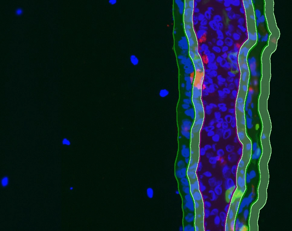
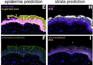
New StrataQuest App: IF ARTIFICIAL SKIN
The IF Artificial Skin App stratifies skin equivalents into dermis and epidermis, further dividing the epidermis into stratum corneum, suprabasal, and basal layers. It outputs area, mean staining intensity, nuclei counts, and % of marker-positive cells for each layer and sublayer. Read publication here
Blog Post
16 Jul, 2024
How Artificial Intelligence Revolutionizes Spatial Phenotyping
Through the power of spatial phenotyping, we gain the ability to shine a spotlight on different cell types and their environments.
Blog Post
06 May, 2024
Imaging in Histopathology: Pyknotic Nuclei
Pyknotic nuclei, key indicators of cell death, are critical in histopathology and disease research. Using advanced imaging and TissueGnostics’ IF Pyknotic Nuclei App, researchers can achieve automated, accurate detection and quantification for deeper diagnostic insights.

Application Note
02 Nov, 2023
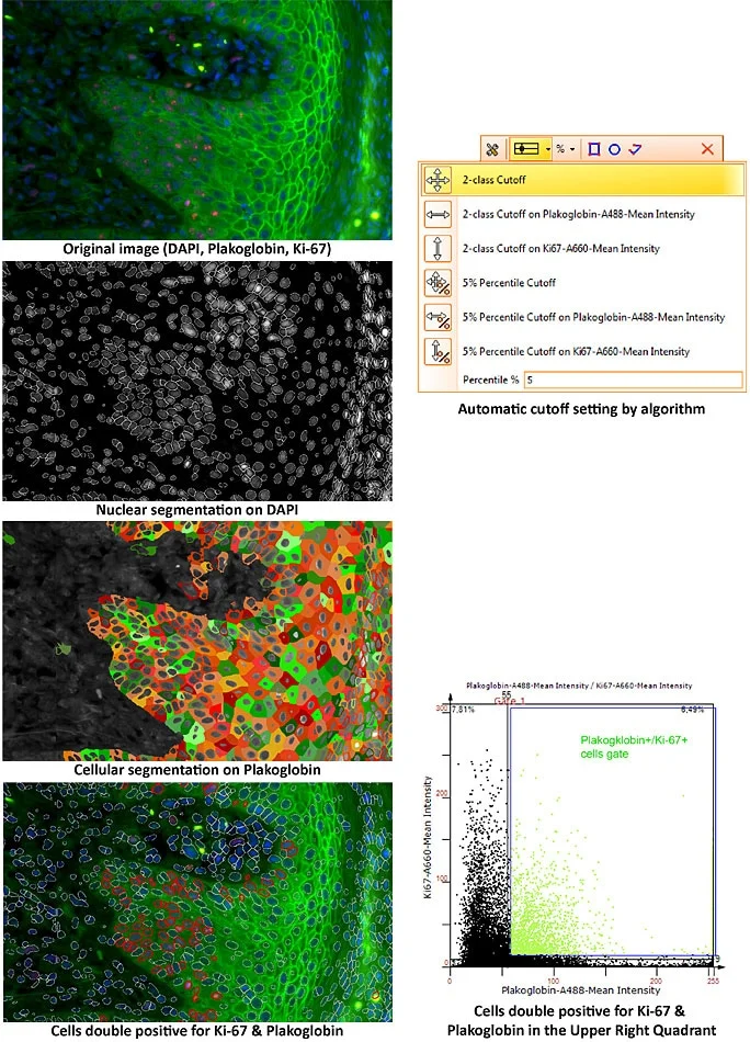
Quantification of Molecular Markers in 2-plex IHC
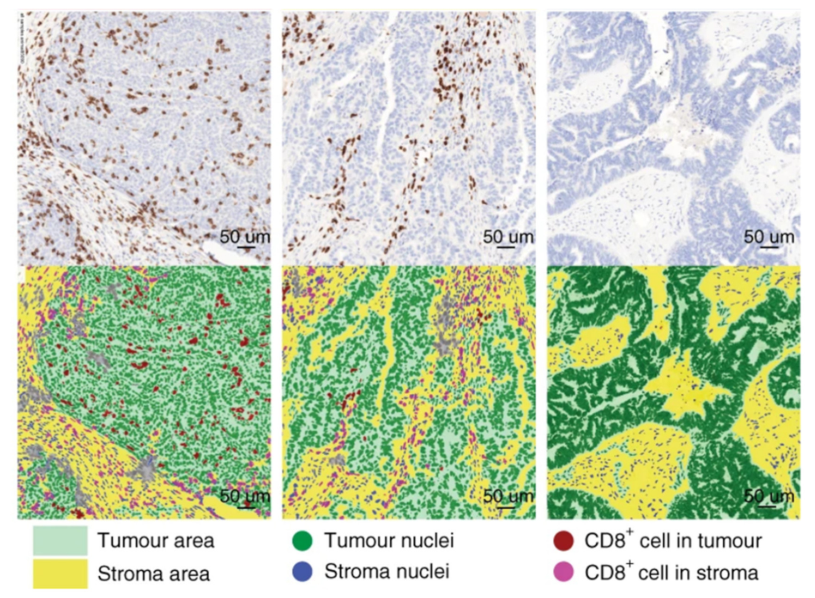
StrataQuest software quantified NCAM and Ki67 distribution in kidney cancer samples, demonstrating how 2-plex IHC staining can be applied to spatial analysis of tumor markers.
Blog Post
09 Oct, 2023
Revolutionizing Tissue Analysis: Image-Based Cytometry
Image-based cytometry is transforming tissue analysis by combining microscopy with flow cytometry features. It enables rapid, automated characterization of cells in their native environment, advancing pathology, oncology, and precision medicine with AI integration.
Blog Post
26 Jun, 2023
Multiplexing Techniques as Useful Tools for High-Content Phenotyping
Multiplexing enables high-content phenotyping by analyzing multiple markers in a single tissue section. Combined with TissueFAXS CHROMA and StrataQuest Apps, it delivers detailed insights into tumor microenvironments and complex cellular interactions.
Blog Post
15 Feb, 2023
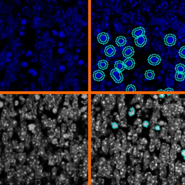
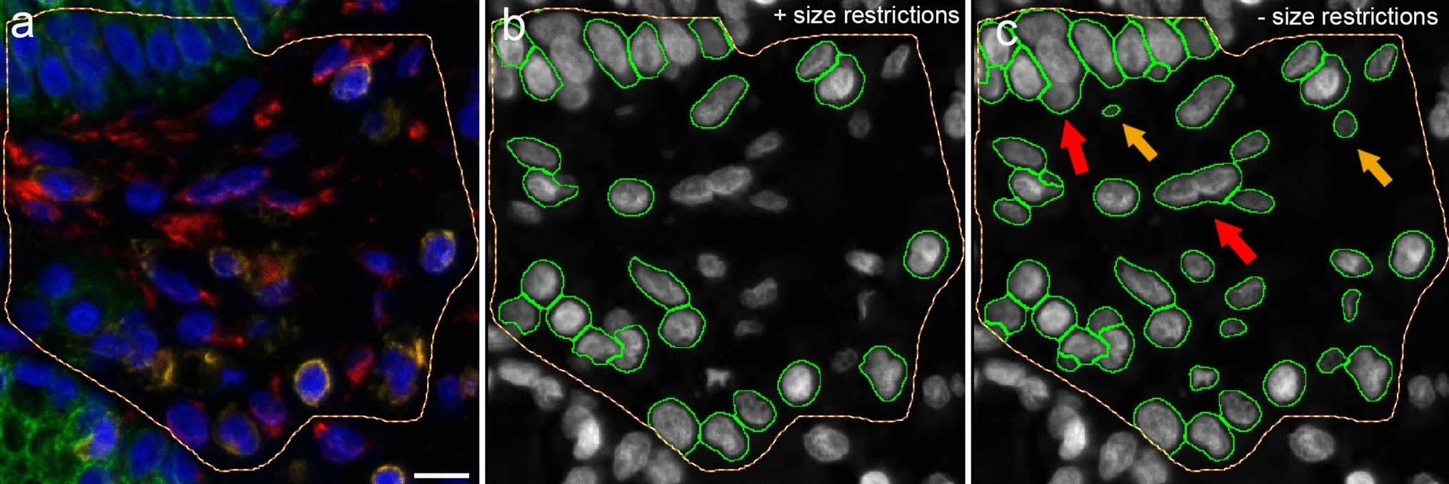
Applications of AI in Cell Segmentation
Accurate cell segmentation is the foundation of tissue cytometry. AI-powered methods, such as deep learning, improve accuracy, reduce errors, and streamline workflows. TissueGnostics integrates AI-based segmentation with phenotyping, spatial analysis, and dot detection.
Blog Post
28 Sep, 2022
What is Imaging Mass Cytometry?
Imaging Mass Cytometry (IMC) expands mass cytometry with laser ablation and metal-tagged antibodies, enabling visualization of 40+ markers in tissue. IMC reveals cellular phenotypes in their native microenvironment, supporting research in oncology, immunology, and precision medicine.
Blog Post
27 Sep, 2022
How Does Imaging Mass Cytometry Work?
Imaging Mass Cytometry (IMC) enables visualization of 40+ markers in tissue using metal-tagged antibodies and mass spectrometry. Combined with StrataQuest analysis, IMC supports single-cell detection, co-expression studies, and spatial mapping for advanced biomedical insights.
Blog Post
01 Sep, 2022
What is Spatial Phenotyping?
Spatial phenotyping combines whole-slide imaging and advanced analysis to study cellular phenotypes in their native context. This approach is key in immunotherapy research, enabling insights into the tumor microenvironment and supporting informed treatment strategies.
Expert Insight
17 May, 2022
The Importance of Immunophenotyping in Research and Pathology
In Trillium Diagnostik, Dr. Felicitas Mungenast and Alexander Stickler-Barang highlight the role of immunophenotyping in tissue sections for diagnosing diseases. The full article is available in German.
White Paper
25 Jan, 2022
PD-L1 raises new hope in the treatment of cutaneous leishmaniasis
PD-L1 expression has emerged as a predictor of treatment response in cutaneous leishmaniasis. Using StrataQuest image analysis, researchers quantified immune markers and parasites, highlighting PD-L1 as a potential host-directed therapeutic target.

Blog Post
07 Dec, 2021
What is Multispectral Cytometry?
Multispectral cytometry resolves spectral overlap to enable accurate, high-parameter analysis of cells and tissues. Learn how this technology, paired with spectral unmixing and advanced imaging, expands applications from cancer research to immunotherapy.
Webinar
03 May, 2021
Automated Imaging of Metabolic Enzyme Activity in the Skin
Prof. Florian Gruber and Christopher Kremslehner (MedUni Vienna) present a metabolic imaging workflow integrating TissueFAXS i PLUS and StrataQuest to study UV-induced effects on skin (Redox Biology).
Customer Publication
28 Apr, 2021
SynNotch-CAR T Cells as Promising Therapy for Solid Tumors
Choe et al. (UCSF) report in Science Translational Medicine on SynNotch-CAR T cells in a glioblastoma PDX model. In-situ T cell dynamics were imaged with TissueFAXS and quantified via StrataQuest.
Customer Publication
01 Mar, 2021
Comprehensive cellular characterization of chronic rhinosinusitis
Giotakis et al. (MedUni Innsbruck) characterized epithelial, connective tissue, and immune cells in chronic rhinosinusitis using TissueFAXS and StrataQuest contextual analysis.
Customer Publication
12 Nov, 2020
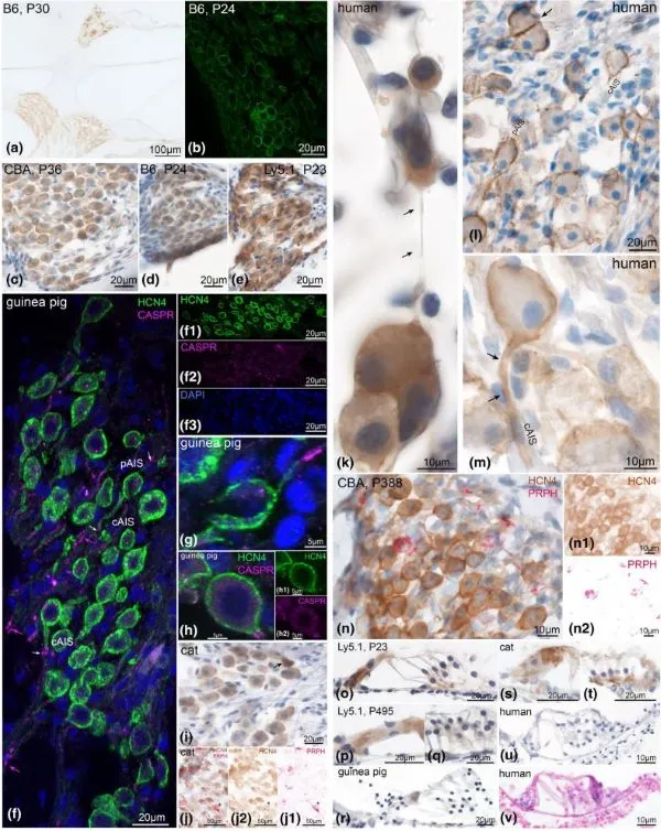
Exploration of neuronal diversity among various species (HCN expression)
A recent publication in the Journal of Neuroscience Research from a research group from Medical University of Innsbruck addresses expression patterns, subcellular location, and age-dependent changes of HCN (voltage gated channel) across different mammalian species. TissueFAXS PLUS and HistoQuest were used to assess expression of HCN in-situ.
