Latest articles in
Blog Post
12 May, 2025
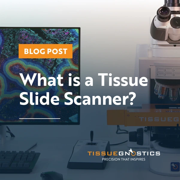
What is a Tissue Slide Scanner?
Tissue slide scanners combine whole-slide imaging, multispectral analysis, and AI-driven software to deliver quantitative, context-rich insights into cells, tissues, and microenvironments, advancing biomedical research.
Blog Post
20 Jan, 2025
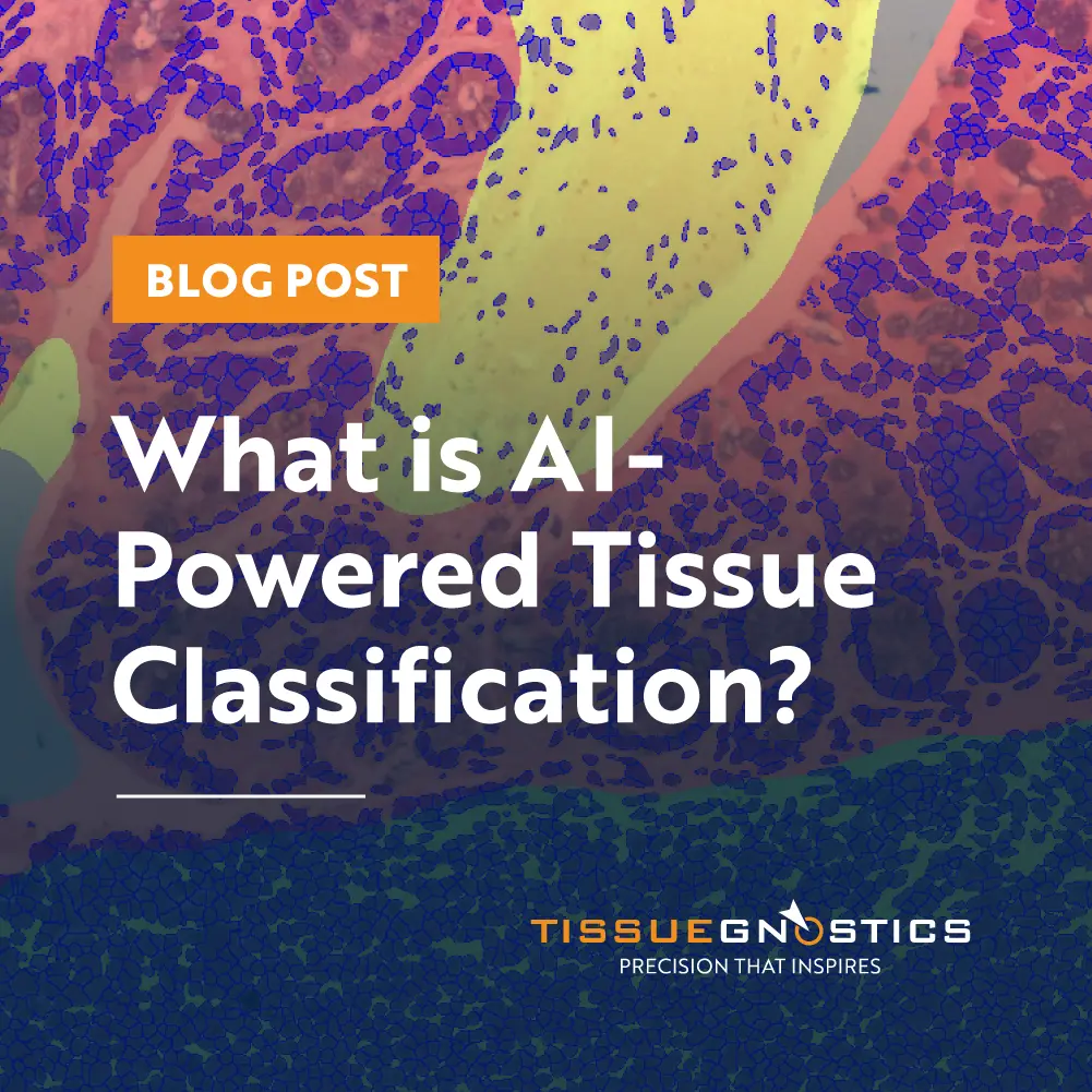
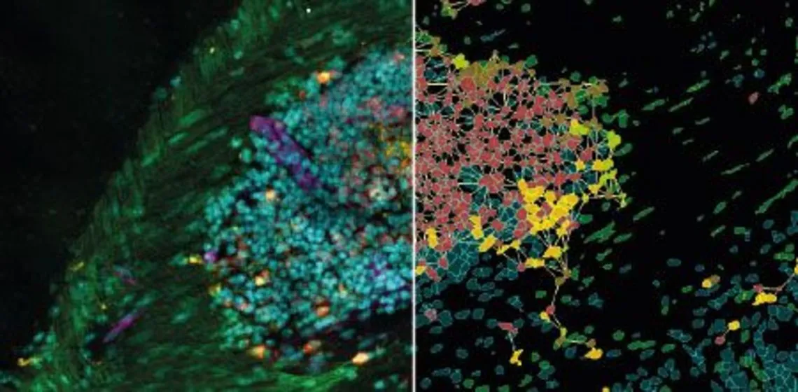
What is AI-Powered Tissue Classification?
AI-powered tissue classification applies machine learning to identify and categorize tissue types. It supports consistent, quantitative analysis of morphology, tumor microenvironments, and disease processes in research and diagnostics.
Blog Post
06 May, 2024
Imaging in Histopathology: Pyknotic Nuclei
Pyknotic nuclei, key indicators of cell death, are critical in histopathology and disease research. Using advanced imaging and TissueGnostics’ IF Pyknotic Nuclei App, researchers can achieve automated, accurate detection and quantification for deeper diagnostic insights.
Blog Post
25 Mar, 2024
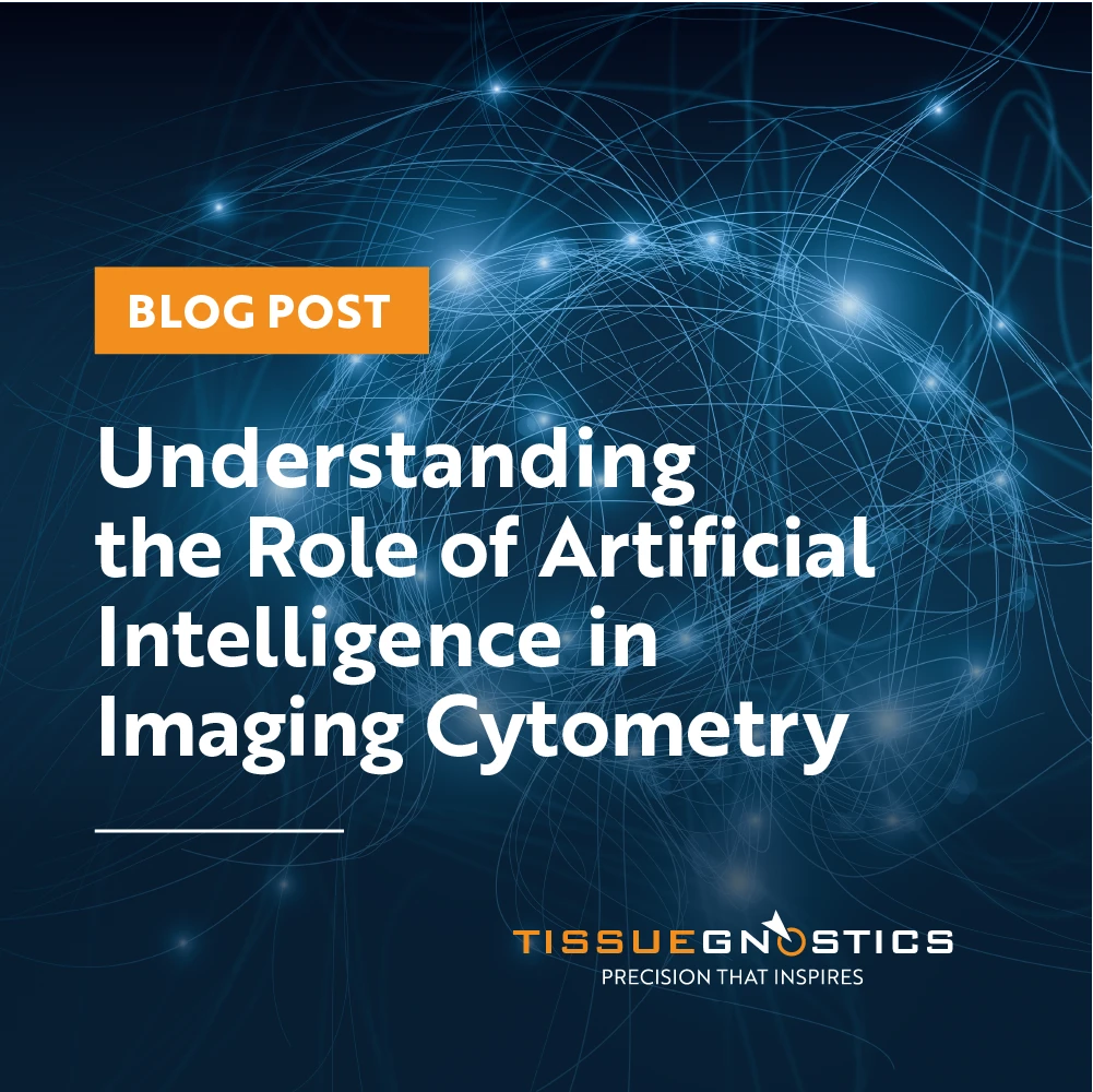
Understanding the Role of Artificial Intelligence in Imaging Cytometry
AI is transforming imaging cytometry by enabling automated, accurate nuclei segmentation and tissue detection. With solutions like StrataQuest, researchers gain higher precision, efficiency, and insights into cellular structures for biomedical research and diagnostics
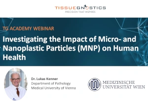
Webinar
19 Mar, 2024
Investigating the Impact of Micro- and Nanoplastic Particles (MNP) on Human Health
Dr. Lukas Kenner, Medical University of Vienna, discusses the impact of micro- and nanoplastic particles on human health, in particular potential effect on cancer development.

Blog Post
08 Nov, 2023
Does Precision Medicine Represent a Healthcare Revolution?
Precision medicine is reshaping healthcare by tailoring treatment to each patient’s molecular profile. With tools like tissue cytometry, TissueFAXS, and StrataQuest, researchers can uncover genetic drivers, track therapy response, and advance personalized care.
White Paper
12 Oct, 2023
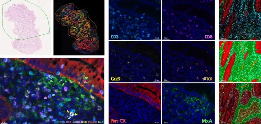
Type I interferon and CD8+ T cell detection predicts response to neoadjuvant treatment of rectal adenocarcinoma
A new study shows that combining type I interferon (MxA) expression with CD8+ T cell density can predict patient response to neoadjuvant treatment in rectal adenocarcinoma. TissueFAXS CHROMA and StrataQuest enabled precise multispectral imaging and analysis.
Blog Post
09 Oct, 2023
Revolutionizing Tissue Analysis: Image-Based Cytometry
Image-based cytometry is transforming tissue analysis by combining microscopy with flow cytometry features. It enables rapid, automated characterization of cells in their native environment, advancing pathology, oncology, and precision medicine with AI integration.
Blog Post
30 Aug, 2023
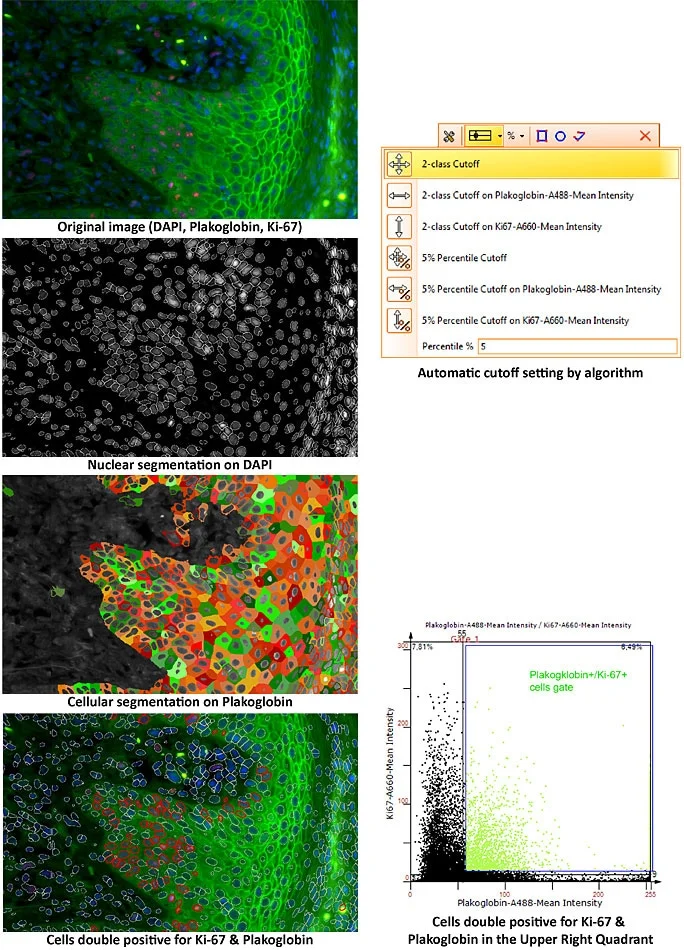
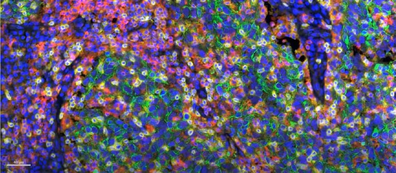
What is Multiplexing & How does it Work?
Multiplexing enables simultaneous detection of multiple markers in a single tissue sample. By combining advanced staining, automated imaging, and AI-powered analysis, it provides deeper insights into the tumor microenvironment and precision medicine.
Expert Insight
24 Apr, 2023
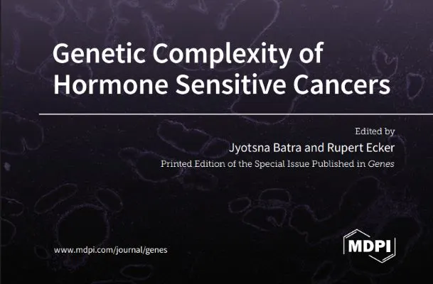
Genetic Complexity of Hormone Sensitive Cancers
A new MDPI Genes special issue, co-authored by Dr. Rupert Ecker (TissueGnostics) & Prof. Jyotsna Batra (QUT), explores genetic mechanisms and digital histopathology in hormone-sensitive cancers. The book covers methods and new insights in cancer biology and is available for free to read.
Application Note
14 Feb, 2023
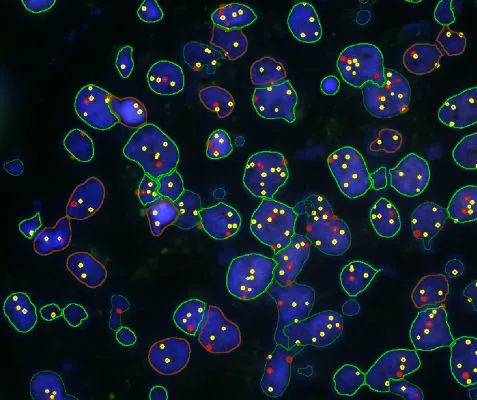
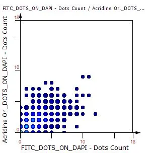
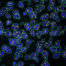
Case Study: Analysis of FISH Using Tissue Cytometry
The IF Dots App in StrataQuest streamlines automated analysis of FISH stainings by detecting nuclei and quantifying dots per cell. This case study shows how the workflow enables accurate, exportable data for genetics research and clinical applications.
Blog Post
06 Feb, 2023
Using FISH Evaluations to Detect Genetic Aberrations in Medicine
FISH enables precise detection of genetic aberrations in cells and is widely applied in diagnostics and personalized medicine. TissueGnostics’ StrataQuest IF Dots App streamlines FISH analysis by automating dot detection and quantification for high-quality data.
Blog Post
18 Jan, 2023
Understanding the Different Types of FISH Evaluations
FISH is a powerful imaging-based technique for detecting DNA and RNA sequences directly in cells and tissues. This post explores the advantages of FISH, its complementary methods like CISH and RNAscope, and how TG solutions streamline automated FISH analysis.
Blog Post
28 Nov, 2022
How FISH Image Analysis Factors into Next-Gen Digital Pathology
How does FISH image analysis advance digital pathology? Learn how whole-slide imaging and AI-powered algorithms enable fast, accurate detection of chromosomal abnormalities, streamline workflows, and open new possibilities for precision medicine.

News
19 Oct, 2022
Centre of Excellence: Medical University Vienna - Institute of Pathophysiology and Allergy Research
The Institute of Pathophysiology and Allergy Research (MedUni Vienna) has been named a Center of Excellence for Quantitative Digital Microscopy by TissueGnostics, recognizing its pioneering use of the TissueFAXS platform in over 45 high-impact publications.

Press Release
27 Sep, 2022
Deep Learning for Oral Dysplasia Classification
TG and the University Clinic of Dentistry (Danube Private University) have received FFG funding for AutOPathStage. This project aims to develop a deep learning–based decision support system for classification of oral dysplasia – a disease of the oral cavity with malignant potential.
Expert Insight
17 May, 2022
The Importance of Immunophenotyping in Research and Pathology
In Trillium Diagnostik, Dr. Felicitas Mungenast and Alexander Stickler-Barang highlight the role of immunophenotyping in tissue sections for diagnosing diseases. The full article is available in German.

Expert Insight
13 Apr, 2022
AI & Healthcare – from Hype to Sustainability: A Global Outlook
As AI moves beyond hype, it is shaping biomedical research and clinical practice. In our Dossier on AI in Biomedicine, experts explore current applications, challenges, and how AI could transform healthcare by 2050.