Nerium Leaf
Segment Nerium leaf into epidermis, hypodermis, mesophyll, and xylem; detect cells, analyze spatial proximity to xylem, and quantify substructure areas and cell distributions.
botanical research
botanical research, botany, plants, nerium leaf, epidermis, hypodermis, mesophyll, xylem
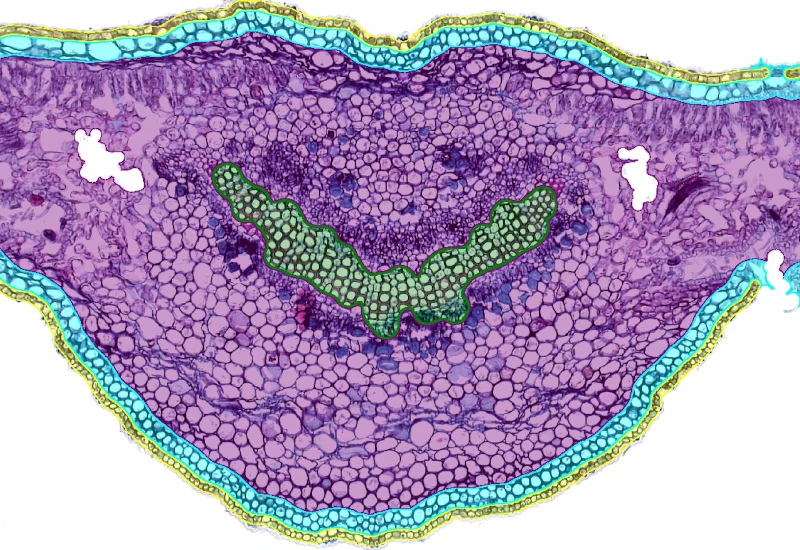
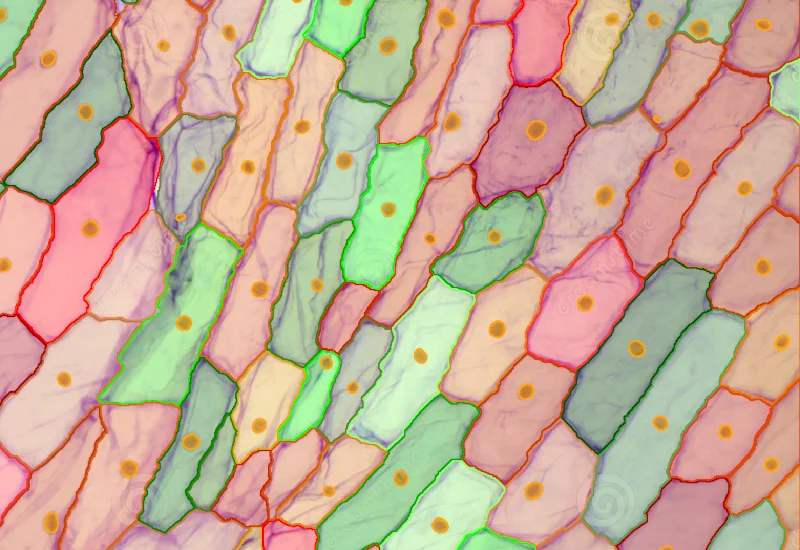
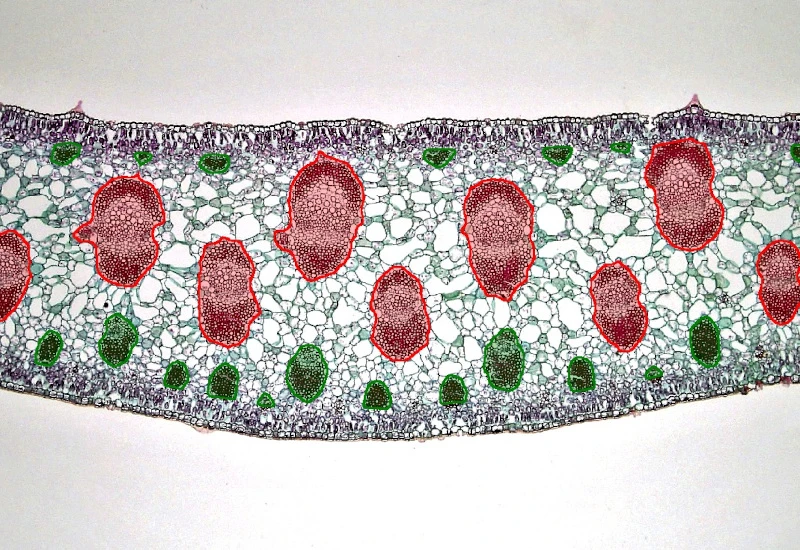
The Nerium Leaf App separates the Nerium leaf into the following anatomical structures: epidermis, hypodermis, mesophyll, and xylem. Additionally, it identifies individual plant cells. It also allows for the spatial identification of cells within user-defined proximities to the xylem substructure. The output includes the area of each anatomical substructure, the total number of cells and the number of identified cells in spatial context
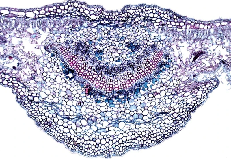
Original Image
Anatomical substructures
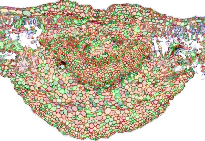
Detection of cells
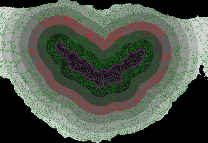
Spatial analysis
botanical research
We support the following file formats:
- TissueFAXS (aqproj)
- StrataFAXS II (vmic)
- PreciPoint (vmic, gtif)
- Generic BigTIFF Import
- Support for multipage BigTIFF files
- OME-TIFF
- JPEG, PNG, BMP, TIFF
- Zeiss (czi)
- Hamamatsu NanoZoomer (ndpi)
- Aperio (svs)
- Leica (scn)
- 3D HISTECH Pannoramic
- Mirax (mrxs)
- Olympus (vsi)
- More slide scanners to be added!
Related Apps
Onion
Quantify onion epidermal cells and nuclei, classify cells into user-defined size ranges, and output total cell number, size (µm²), density, and counts per size category.
botanical research, botany, plants, onion, epidermal cells

Plant Root
Segment plant roots into epidermis, cortex, endodermis, pericycle, and stele; detect cells and quantify area and cell count per anatomical structure.
botanical research, botany, plants, plant root, epidermis, cortex, endodermis, pericycle, vascular cylinder
Yucca Leaf
Segment Yucca leaf into epidermis, collenchyma, parenchyma, and vascular bundles; detect cells, classify vascular bundles by size, and quantify substructure areas and spatially defined cell distributions.
botanical research, botany, plants, yucca leaf, epidermis, collenchyma, parenchyma, vascular bundles

Custom App development
Perfectly tailored image analysis solutions for your research.
You have a specific research question that needs to be answered? We offer custom development of image analysis pipelines for specific tasks, be it detection of cellular phenotypes or quantification of tissue structures. After discussing your goals with one of our experts, you will get a ready-to-use App and be a step closer to an impactful publication.