IHC Necrotic Tumor
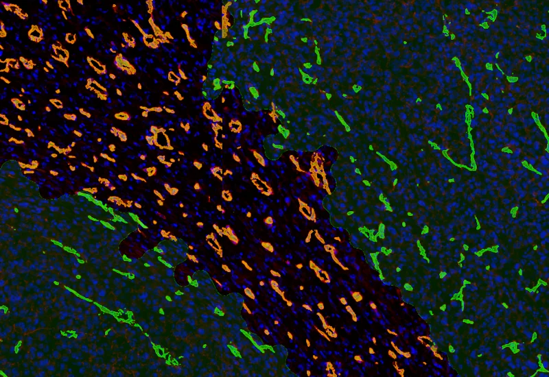
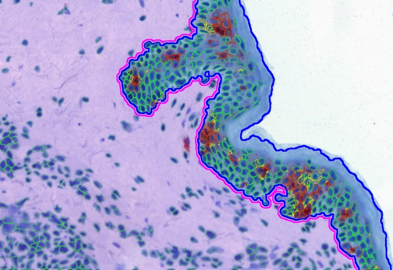
AI-based segmentation of tumor tissue into tumor, necrotic tissue, and stroma with neutrophil detection and compartment-based quantification.
metastructures
single-cell analysis
tumor microenvironment
immunophenotyping
neutrophiles, tumor, necrosis, necrotic tissue, immunophenotyping, mouse tissue
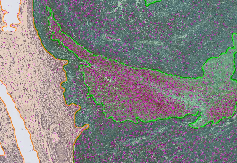
The IHC Necrotic Tumor App is able to segment tumor tissues into tumor, necrotic tissue and stroma using the AI Classifier. Furthermore, it identifies single cells as well as one additional cellular marker (e.g. Neutrophils). It outputs area of tumor, necrotic tissue and stroma. Further measurements are the number and percentage of neutrophils within the morphological entities.

Original Image
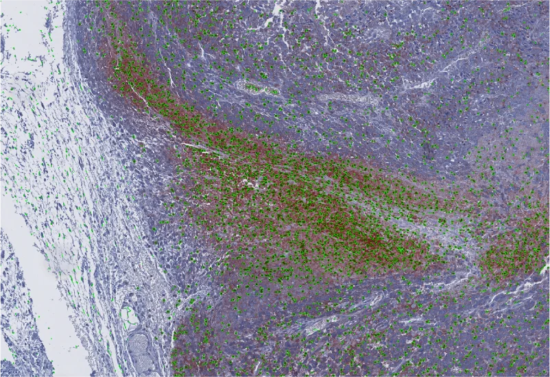
Marker-positive cell detection
Stroma/Tumor/Necrotic tissue detection
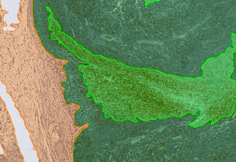
Combined detection
immunophenotyping
Webinar
20 Jan, 2026
Decoding Metastatic Potential in Colorectal Cancer Using Tissue Cytometry
tumor microenvironment
Blog Post
07 Apr, 2026
How immunofluorescence image analysis factors into NSCLC studies
single-cell analysis
White Paper
30 Mar, 2026
Understanding NeuroCOVID-19: SARS-CoV-2 Disrupts Astrocyte Homeostatic Functions
metastructures

Blog Post
17 May, 2023
An Intro to Deep Learning in Biomedical Imaging
We support the following file formats:
- TissueFAXS (aqproj)
- StrataFAXS II (vmic)
- PreciPoint (vmic, gtif)
- Generic BigTIFF Import
- Support for multipage BigTIFF files
- OME-TIFF
- JPEG, PNG, BMP, TIFF
- Zeiss (czi)
- Hamamatsu NanoZoomer (ndpi)
- Aperio (svs)
- Leica (scn)
- 3D HISTECH Pannoramic
- Mirax (mrxs)
- Olympus (vsi)
- More slide scanners to be added!
Related Apps
IF Tumor Vascularization
Segment tissue into tumor and stroma/healthy areas, detect CD31+ vessels, and quantify vessel number, area, density, and connectivity with configurable wall closing and distance linking.
vasculatization, cancer, stroma, tumor, blood vessels, CD31, spatial analysis, tumor microenvironment
IHC Macrophages
Detect macrophages in IHC samples, apply area and distance-range algorithms to measure Langerhans cell distances to the epidermis border inside/outside, and export up to 20 parameters per cell compartment plus boundary distance.
immune cells, brightfield, macrophages, immunohistochemistry, epidermis, langerhans cells, dermatology, CD68

IHC Necrotic Tumor Angio
Segment tumor tissue into tumor, necrotic tissue, and blood vessels using an AI classifier, and quantify compartment areas, total vessel count, and vessel distribution across morphological entities.
tumor immune microenvironment, tumor microenvironment, necrosis, blood vessles, vascularization, tumor

Custom App development
Perfectly tailored image analysis solutions for your research.
You have a specific research question that needs to be answered? We offer custom development of image analysis pipelines for specific tasks, be it detection of cellular phenotypes or quantification of tissue structures. After discussing your goals with one of our experts, you will get a ready-to-use App and be a step closer to an impactful publication.