
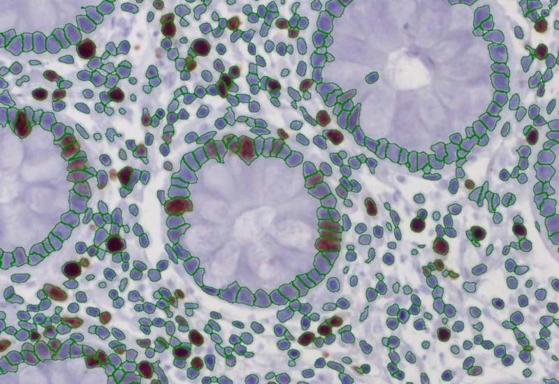
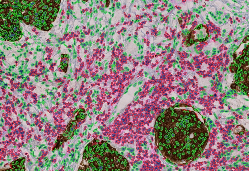
IHC 2
Separate two markers in IHC/HC slides, segment single cells into nucleus, perinuclear area, and/or cytoplasm, measure up to 20 intensity, statistic, and morphometric parameters per compartment.
single-cell analysis
cell culture
immunophenotyping
immunohistochemistry, phenotypes, biomarker, brightfield
The IHC 2 App separates two markers (e.g. chromogen and counterstain) in an IHC or HC digital slide and segments single cells into nucleus, and/or perinuclear area and/or cytoplasm. Each segmented cell compartment is measured for up to 20 intensity, statistic and morphometric parameters which are displayed in scattergrams and histograms and can be exported.

Original image
Nuclei detection
Phenotype detection
immunophenotyping
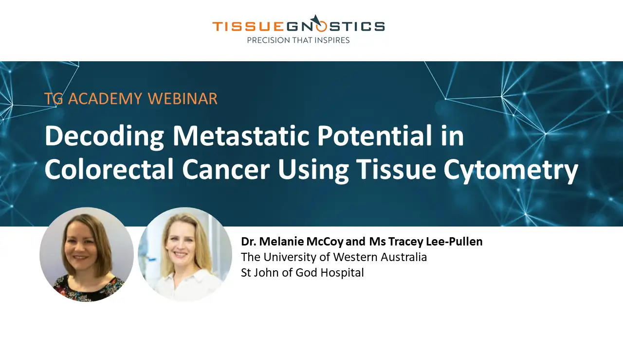
Webinar
20 Jan, 2026
Decoding Metastatic Potential in Colorectal Cancer Using Tissue Cytometry
cell culture

Webinar
19 Nov, 2024
Quantification of p-H2AX Foci in Co-cultured Cells Exposed to Radiation, Livia Sima
single-cell analysis
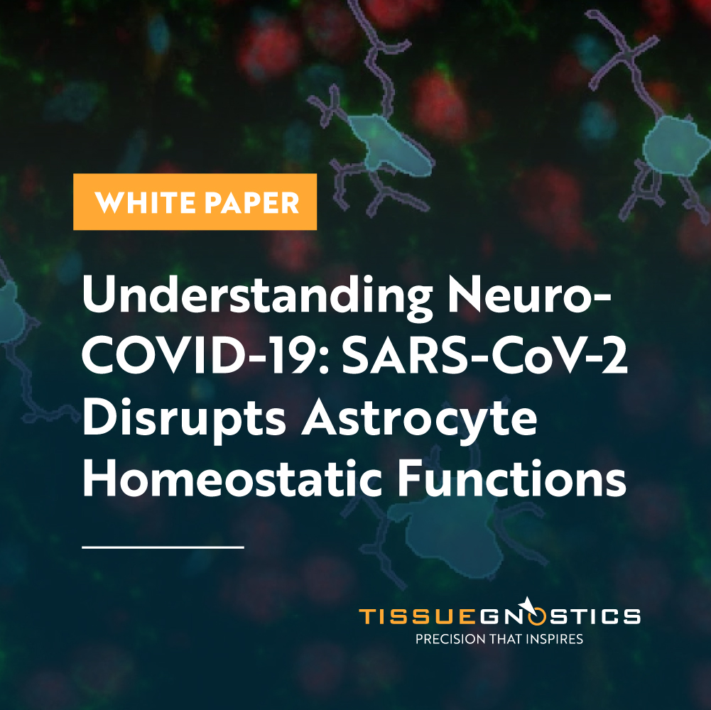
White Paper
30 Mar, 2026
Understanding NeuroCOVID-19: SARS-CoV-2 Disrupts Astrocyte Homeostatic Functions
We support the following file formats:
- TissueFAXS (aqproj)
- StrataFAXS II (vmic)
- PreciPoint (vmic, gtif)
- Generic BigTIFF Import
- Support for multipage BigTIFF files
- OME-TIFF
- JPEG, PNG, BMP, TIFF
- Zeiss (czi)
- Hamamatsu NanoZoomer (ndpi)
- Aperio (svs)
- Leica (scn)
- 3D HISTECH Pannoramic
- Mirax (mrxs)
- Olympus (vsi)
- More slide scanners to be added!
Related Apps
IHC Adipocyte
The IHC Adipocyte App quantifies adipocytes and their lumina in HE samples, correcting membrane artifacts and providing area measurements.
adipocytes, fat tissue, fat cells, H&E
IHC 3
Unmix three markers in IHC/HC slides, segment single cells into nucleus, perinuclear area, and/or cytoplasm, measure up to 20 intensity, statistic, and morphometric parameters per compartment.
immunohistochemistry, brightfield

Custom App development
Perfectly tailored image analysis solutions for your research.
You have a specific research question that needs to be answered? We offer custom development of image analysis pipelines for specific tasks, be it detection of cellular phenotypes or quantification of tissue structures. After discussing your goals with one of our experts, you will get a ready-to-use App and be a step closer to an impactful publication.