
IF Spheroids
Detect spheroids and nuclei-based cells, segment cellular compartments, quantify multiple IF markers and dot signals, and analyze spatial proximity of cell populations within spheroids.
single-cell analysis
dot detection
spheroid, membrane, 2D and 3D culture, Human colon cancer cell lines
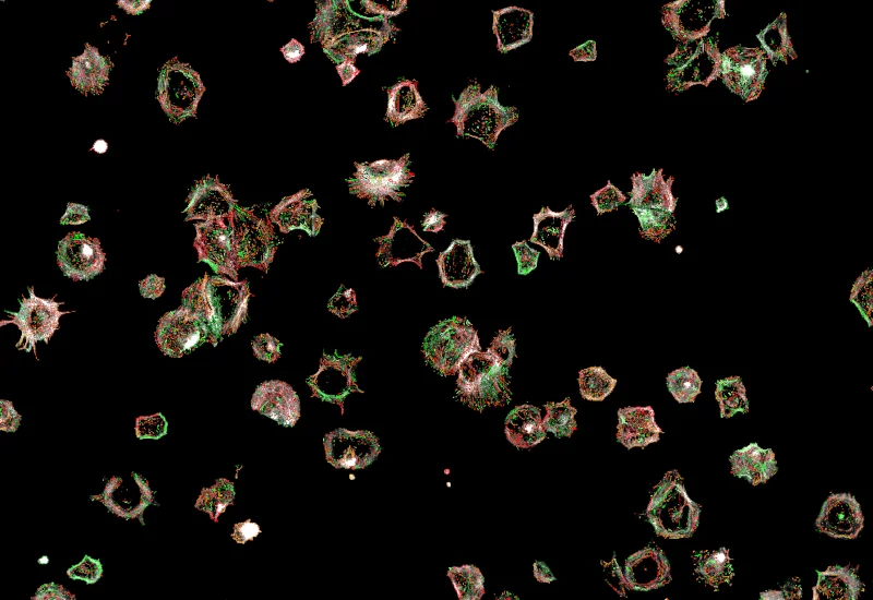
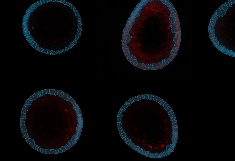
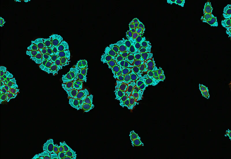
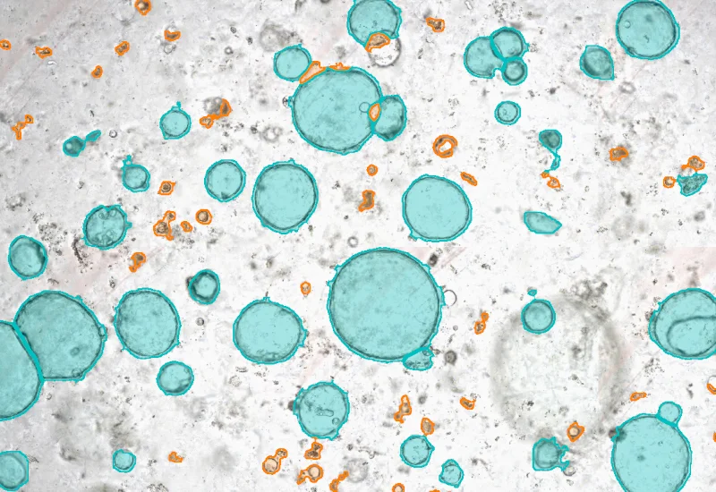
The IF Spheroids App allows a comprehensive analysis of spheroids (as wells organoids and embryoid bodies). It automatically identifies the spheroids and the cells based on the nuclei staining. It analyzes 2 additional IF markers. It segments the cells into different cellular compartments including membrane, nuclei and cytosol and further measures the marker expression for each compartment. It also can measure dot markers (if available). It established proximity distances for the cells detected within the sperhoids and thereby brings the IF stained cell populations into their spatial relationships.
Riedl A, Schlederer M, Pudelko K, et al. Comparison of cancer cells in 2D vs 3D culture reveals differences in AKT-mTOR-S6K signaling and drug responses. J Cell Sci. 2017;130(1):203-218. doi:10.1242/jcs.188102
Image courtesy of Michaela Schlederer, Medical University of Vienna, Austria
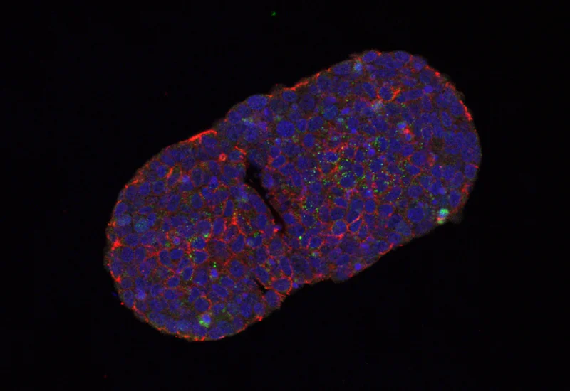
Original Image
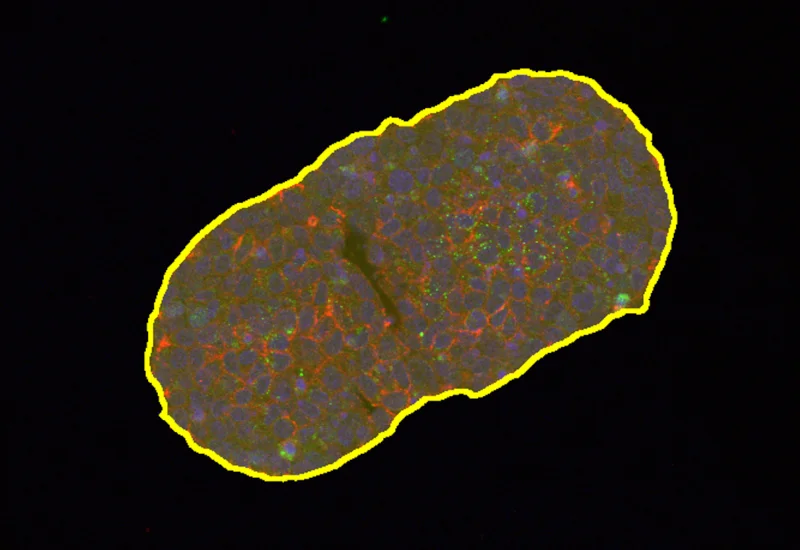
Spheroid/Organoid detection
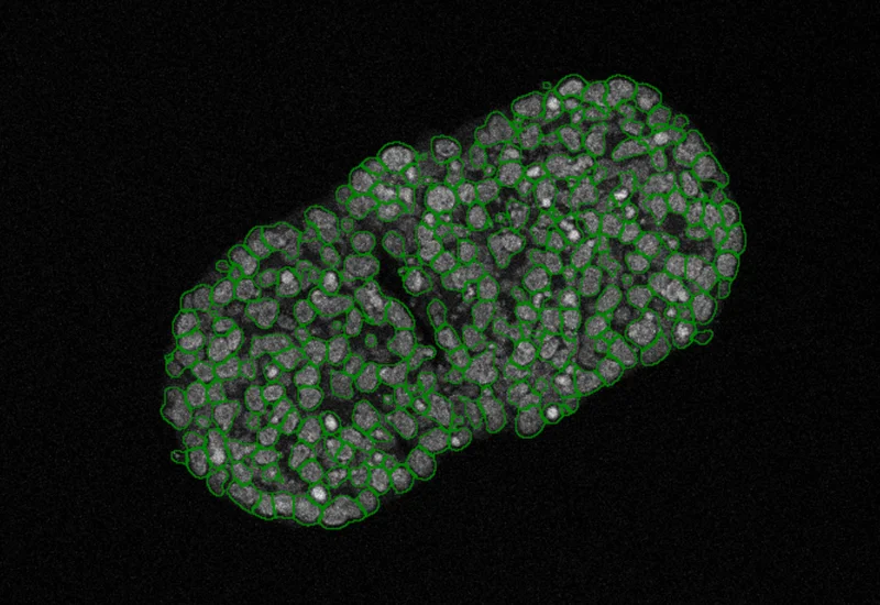
Nuclei detection
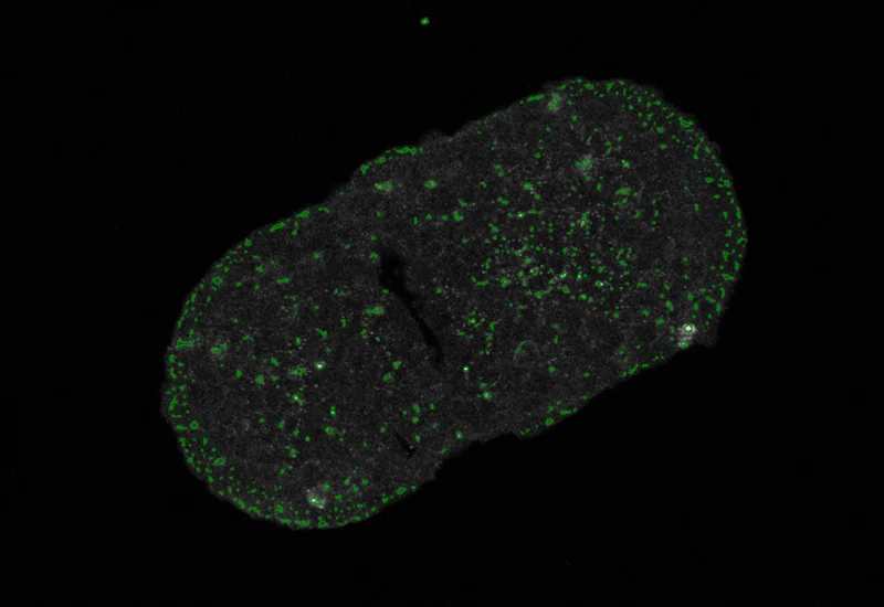
Dot detection

Nuclei/cytoplasm/membrane detection
Proximity measurement
dot detection
Blog Post
15 Feb, 2023
Applications of AI in Cell Segmentation
single-cell analysis
White Paper
30 Mar, 2026
Understanding NeuroCOVID-19: SARS-CoV-2 Disrupts Astrocyte Homeostatic Functions
We support the following file formats:
- TissueFAXS (aqproj)
- StrataFAXS II (vmic)
- PreciPoint (vmic, gtif)
- Generic BigTIFF Import
- Support for multipage BigTIFF files
- OME-TIFF
- JPEG, PNG, BMP, TIFF
- Zeiss (czi)
- Hamamatsu NanoZoomer (ndpi)
- Aperio (svs)
- Leica (scn)
- 3D HISTECH Pannoramic
- Mirax (mrxs)
- Olympus (vsi)
- More slide scanners to be added!
Related Apps
IF Embryoid Bodies
Detect embryoid bodies/organoids in IF-stained samples, segment nuclei, identify cellular phenotypes in nuclei or membrane, and quantify organoid number, area, and phenotype distribution.
embryoid bodies, cell culture

Custom App development
Perfectly tailored image analysis solutions for your research.
You have a specific research question that needs to be answered? We offer custom development of image analysis pipelines for specific tasks, be it detection of cellular phenotypes or quantification of tissue structures. After discussing your goals with one of our experts, you will get a ready-to-use App and be a step closer to an impactful publication.