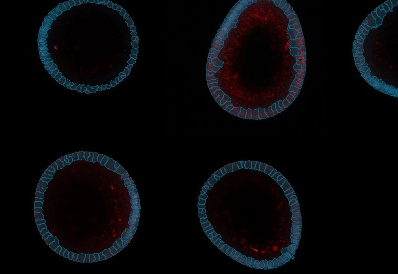

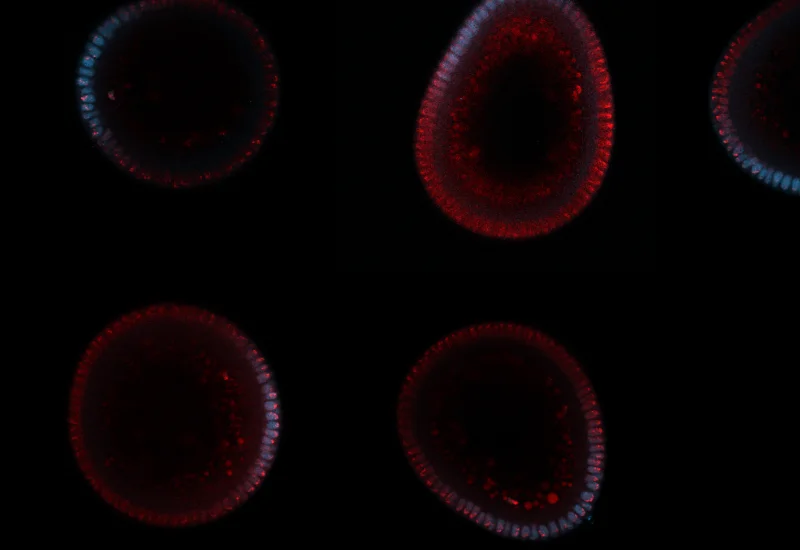
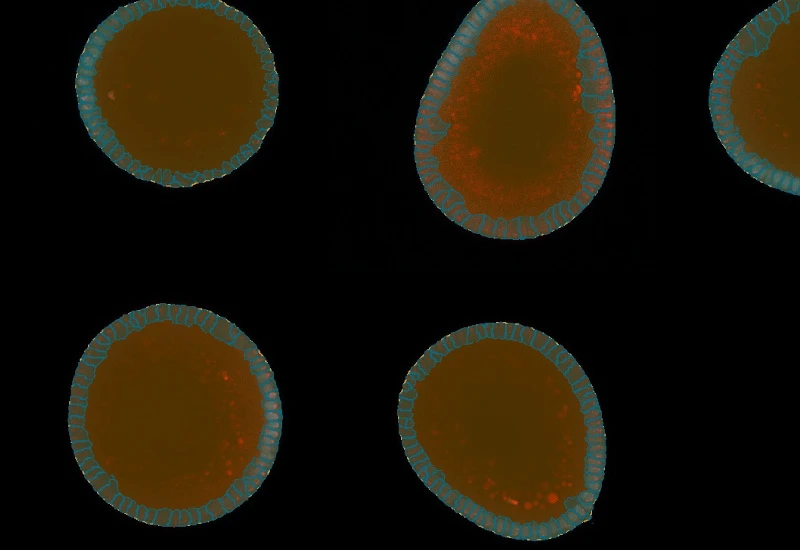
IF Embryoid Bodies
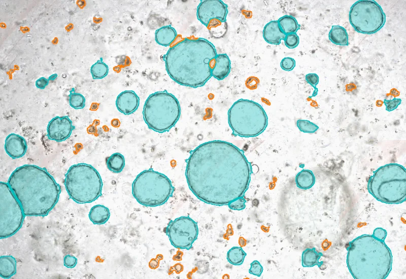
Detect embryoid bodies/organoids in IF-stained samples, segment nuclei, identify cellular phenotypes in nuclei or membrane, and quantify organoid number, area, and phenotype distribution.
single-cell analysis
cell culture
organoid detection
embryoid bodies, cell culture
The IF Embryoid Bodies App automatically detects embryoid bodies/organoids based on IF staining. It identifies nuclei based on DAPI staining or other nuclei dye and identifies additional phenotype markers in the nuclei/cell and/or membrane of the detected cells. It outputs number and area (µm2) of detected embryoid bodies/organoids, count of nuclei and count/% of cellular phenotypes.
organoid detection
Webinar
26 May, 2025
Exploring Immune Cell Interactions and Tissue Regeneration through Imaging
cell culture

Webinar
19 Nov, 2024
Quantification of p-H2AX Foci in Co-cultured Cells Exposed to Radiation, Livia Sima
single-cell analysis
White Paper
30 Mar, 2026
Understanding NeuroCOVID-19: SARS-CoV-2 Disrupts Astrocyte Homeostatic Functions
We support the following file formats:
- TissueFAXS (aqproj)
- StrataFAXS II (vmic)
- PreciPoint (vmic, gtif)
- Generic BigTIFF Import
- Support for multipage BigTIFF files
- OME-TIFF
- JPEG, PNG, BMP, TIFF
- Zeiss (czi)
- Hamamatsu NanoZoomer (ndpi)
- Aperio (svs)
- Leica (scn)
- 3D HISTECH Pannoramic
- Mirax (mrxs)
- Olympus (vsi)
- More slide scanners to be added!
Related Apps

IF Dots
Detect dots-stainings per cell in up to four markers, segment cellular compartments, measure up to 20 intensity, statistic, and morphometric parameters, and quantify dot count, mean intensity, total area, intensity sum, and per-dot area/intensity per compartment.
cell culture, breast cancer, fluorescence, HER2


Custom App development
Perfectly tailored image analysis solutions for your research.
You have a specific research question that needs to be answered? We offer custom development of image analysis pipelines for specific tasks, be it detection of cellular phenotypes or quantification of tissue structures. After discussing your goals with one of our experts, you will get a ready-to-use App and be a step closer to an impactful publication.