Endometrium Morphology
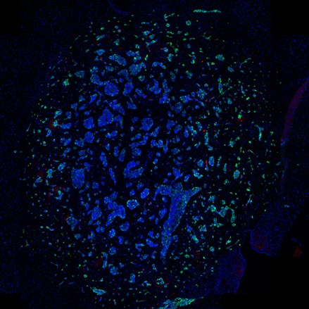
Segment H&E-stained endometrium into glands, stroma, and blood vessels, and quantify the area of each morphological entity.
metastructures
tumor microenvironment
vascularization
H&E
endometrium, endometriosis, H&E, glands, blood vessels, stroma, gynecology
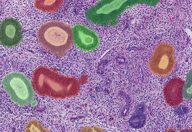
The Endometrium HE App allows the segmentation of hematoxylin and eosin stained endometrium tissues into their morphological entities including glands, stroma and blood vessels.The measurements provided by the App are area of glands, stroma and blood vessels.
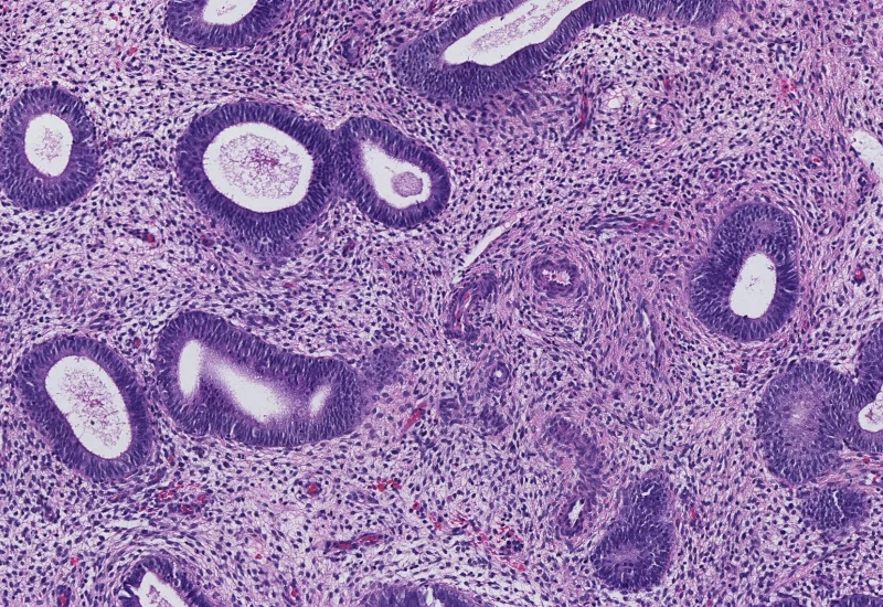
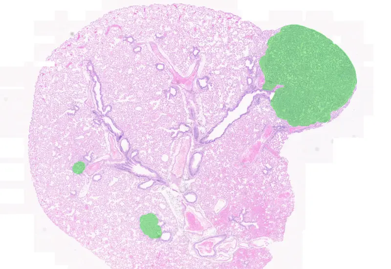
Original Image
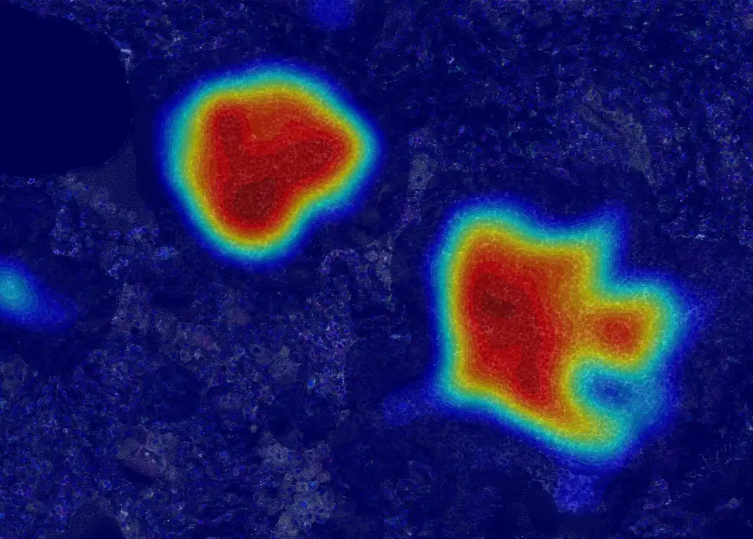
Gland detection
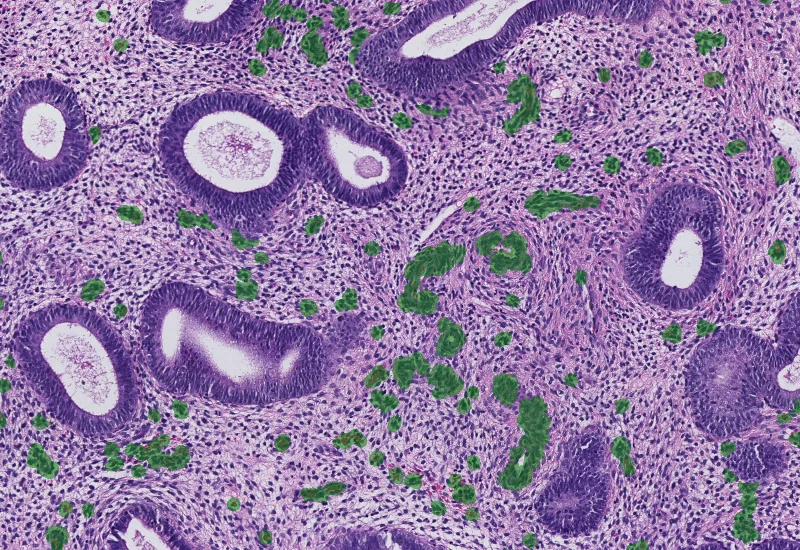
Vessel detection
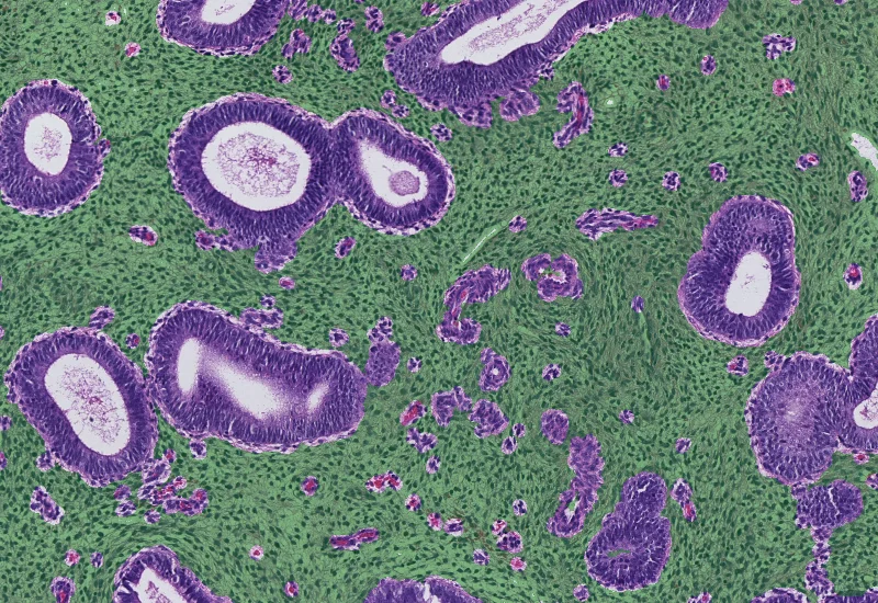
Stroma detection

Combined detection
H&E
vascularization
Application Note
01 Jun, 2023
Evaluating the Distance of Tumor Cells from Blood Vessels
tumor microenvironment
Application Note
06 Jul, 2026
Mapping spatial cell-to-cell interactions in tertiary lymphoid structures with spatial proteomics
metastructures

Blog Post
17 May, 2023
An Intro to Deep Learning in Biomedical Imaging
We support the following file formats:
- TissueFAXS (aqproj)
- StrataFAXS II (vmic)
- PreciPoint (vmic, gtif)
- Generic BigTIFF Import
- Support for multipage BigTIFF files
- OME-TIFF
- JPEG, PNG, BMP, TIFF
- Zeiss (czi)
- Hamamatsu NanoZoomer (ndpi)
- Aperio (svs)
- Leica (scn)
- 3D HISTECH Pannoramic
- Mirax (mrxs)
- Olympus (vsi)
- More slide scanners to be added!
Related Apps
Tumor Foci
Detect whole tissue and tumor foci based on using nuclear structure analysis, measure number of tumor foci, area, and cellular density.
H&E, cancer tissue, TMA

Wilms Tumor
AI-based segmentation of H&E-stained Wilms tumor sections into tumor, stroma, and blood vessels with quantitative area measurements.
wilms tumor, H&E, nephroblastoma, kidney, renal cancer
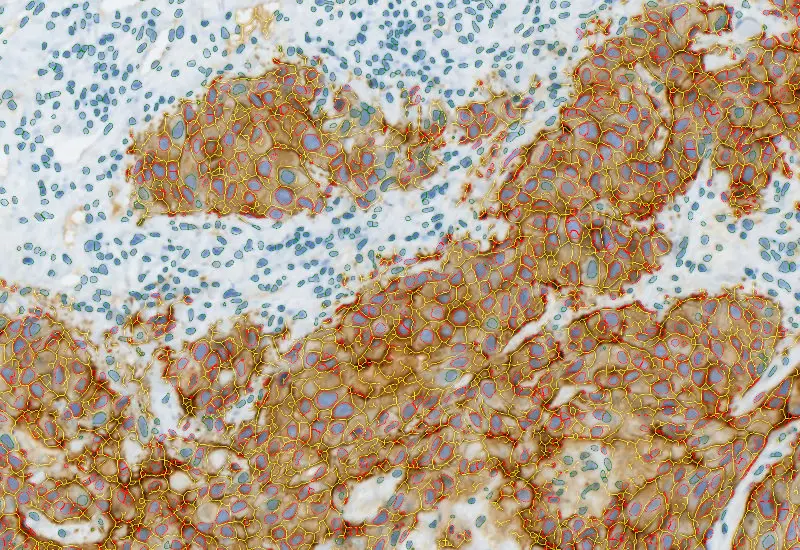
IHC Membrane
Unmix up to three markers in IHC/HC slides, segment cells into nucleus, perinuclear area, cytoplasm, and membrane (e.g. HER2/neu), measure up to 20 intensity, statistic, and morphometric parameters per compartment plus membrane intensity and angle.
HER2, breast cancer, immunohistochemistry, automated scoring

Custom App development
Perfectly tailored image analysis solutions for your research.
You have a specific research question that needs to be answered? We offer custom development of image analysis pipelines for specific tasks, be it detection of cellular phenotypes or quantification of tissue structures. After discussing your goals with one of our experts, you will get a ready-to-use App and be a step closer to an impactful publication.