Striosomes Causal Changes in Task Engagement
Febuary, 2021
Learning through affective responses, in which experiences are regarded as having a positive or negative valence (e.g., pleasure or fear, respectively), requires a judgment of the relative value of options. Several studies have linked a cluster of neurons in the subcortical basal ganglia, known as the striatum, to learning these valence-based responses (1,2)
. As a key part of the brain's movement and motivation control system, the striatum is associated with a range of neurological diseases, such as Parkinson's disease and Huntington's disease (HD). Researchers are just beginning to unravel the complexity of the striatum, which is composed of multiple neuronal phenotypes and organized into neurochemically distinct compartments. These compartments present as pockets of striosomes embedded in a larger surrounding matrix compartment.
Striosomes Mediate Value-Based Learning Vulnerable in Age and a Huntington's Disease Model
Studies that have implicated striosomes in reinforcement-related updating paradigms and cost-benefit conflict decision-making have used well-trained subjects. As such, it is unclear how striosomes perform during the naïve process of learning decision-making under potentially rewarding and costly outcomes. A research team from the Massachusetts Institute of Technology, Harvard, and Stanford University investigated the neurobiology of decision-making during valence-discrimination learning across healthy aging and in a murine model of HD and tracked the activity of striatal microcircuits interconnecting striosomes. Friedman et al. found that the learning signals in striosomes scale according to subjective value and decline during aging and in neurodegenerative disorders. This study offers evidence of a mechanistic cause to explain how the motivation to learn new tasks and maintain engagement is negatively correlated with age.
Establishing Correlation
Through a series of experiments, mice were played to two different tones; one was coupled with a rewarding delivery of sucrose and the other was coupled with an aversive bright light. Through daily training, mice learned that increased licking of a spout upon hearing the first tone would result in more reward, whereas reduced licking during the second tone would result in receiving a less aversive light intensity. As mice learned this task, the researchers observed that striosomes signals underwent selective modification, such that their frequency would decline while their amplitude increased. The same activity in the matrix was not recorded. This suggested that striosomal activity is shaped by discrimination learning. Subsequent experimentation revealed that striosomes were predominantly responsible for encoding discrimination levels during learning and that striosomal signals — and not matrix signals — are sensitive to task engagement and correspond to subjective value.
Establishing Causation
To investigate causality, the researchers employed chemogenetic methods. By selectively expressing designer receptors exclusively activated by designer drugs (DREADDS) in striosomal and matrix neurons, they were able to modulate specific neuronal activity through the DREADDS ligand, clozapine-N-oxide (CNO). By stimulating inhibitory DREADDS on striosomes and so reducing striosomal activity (as measured through Ca2+ transients), mice exhibited a decrease in task engagement. No such effect was observed following the same inhibitory manipulation in matrix populations. Conversely, stimulation of excitatory DREADDS on striosomes led to greater task engagement. As before, the same effect was no observed following stimulation of excitatory matrix populations. Altogether, the researchers were able to demonstrate that striosomal populations, and not matrix populations, drive task engagement.
TissueGnostics
TissueGnostics, a solution provider for Precision Medicine / Next-Generation Digital Pathology, supported the experiments outlined above through its fully integrated, cutting-edge tissue cytometers.
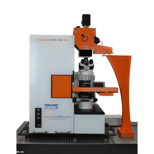
Staining was performed on tissue from the offspring, to tag and characterize striatal compartments following the generation of mouse lines (D1/D2-GFP & Mash1/Dlx1). Quadruple protein stains (VGLUT1/GFP/PV/MOR1), and triple protein stains (mHTT/GFP/PV and GFP/mCherry/MOR1) were captured using TissueGnostics': TissueFAXS SL Q, the high-end confocal Slide Loader system (automatic acqusition of up to 120 slides). This system uses TissueGnostics' spinning disc confocal technology with a slide autoloader for high-throughput, tiled confocal imaging. Initial imaging of the dorsal central striatum allowed the researchers to trace and select regions of interest for acquisition.
Moreover, by using the extended focus option, they were able to automatically calculation a maximum projection. Following the acquisition, images were validated for focus quality. TissueGnostics Slide Validator technology ensures a high level of digital slide sharpness. Each scanned field of view (FOV) is evaluated for sharpness on-the-fly during the scan process. If the validation algorithm deems the FOV insufficiently sharp it will be rescanned on-the-fly.
Also, all staining conducted in the DREADD experiments collected images using the TissueFAXS Whole Slide Scanning System, which is available through the TissueFAXS system. This system is a whole slide imaging tissue cytometer with multichannel fluorescence and brightfield imaging capability. High-throughput, tiled epifluorescence imaging was achieved using the motorized stage and autofocus algorithm. Using the standard configuration — an 8 slide capacity — the researchers produced a 2.5x preview scan of all slides. This allowed them to trace and select regions of interest in the dorsal central striatum for acquisition in higher resolution. Once acquisition finished, images were validated for focus quality, as above.
TissueGnostics’ instruments and technology-facilitated Friedman et al. in drawing causal links between striosomal activity modulating task engagement, and not matrix activity, through DREADDS. While this experiment demonstrates the utility of TissueGnostics instruments and technology, TissueGnostics offers a broader range of products covering an even wider variety of applications.
References and Further Reading
1. Berridge, K.C. (2019) Affective valence in the brain: modules or modes? Nature Reviews Neuroscience, 20 (4): 225–234. doi:10.1117/12.2549369.Hyperspectral.
2. Perosa, V., De Boer, L., Ziegler, G., et al. (2020) The Role of the Striatum in Learning to Orthogonalize Action and Valence: A Combined PET and 7 T MRI Aging Study. Cerebral Cortex, 00 (00): 1–12. doi:10.1093/cercor/bhz313.
This article is based on the paper: Friedman, A., Hueske, E., Drammis, S.M., et al. (2020) Striosomes Mediate Value-Based Learning Vulnerable in Age and a Huntington's Disease Model. Cell, 183 (4): 918-934.e49. doi:10.1016/j.cell.2020.09.060.
Click here to read the full paper!