Department for Immunology, Leibniz Institute for Immunotherapy (LIT)
The Department for Immunology at the LIT – Leibniz Institute for Immunotherapy in Regensburg, Germany, is recognized as a TGCenter of Excellence for its long-standing collaboration since 2012. Led by Prof. Uwe Ritter, the team has made significant scientific contributions through numerous high-impact publications using TissueGnostics technologies. Their continued participation in joint webinars and conference activities exemplify a highly impactful partnership. »Interview with Prof. Uwe Ritter
Interview with Prof. Dr. Uwe Ritter
Department for Immunology, Leibniz Institute for Immunotherapy (LIT), Regensburg, Germany Center of Excellence award ceremony. From left to right, Rudolf Jedletzberger (COO of TissueGnostics), Rupert Ecker (CEO of TissueGnostics) and Uwe Ritter.
Briefly describe your research interestsand role at the Department of Immunology at Leibniz Institute for Immunotherapy (LIT).
I am working as a principal investigator and group leader at the Leibniz Institute for Immunotherapy (LIT) and am interested in the so-called skin-associated lymphatic tissue (SALT).
Me and my group are investigating how antigen-specific immune responses are induced and regulated in skin-draining lymph nodes. In this context we are focused on myeloid cells and adaptive immune responses, resulting from interactions between antigen-presenting cells and T cells.
Currently, we are characterizing the cellular checkpoints resulting in the initiation and regulation of T cell-mediated immunity. The impact of myeloid subsets in immune regulation and wound healing is of special interest. Our goal is the identification of exo- and endogenous mediators that are interfering with checkpoints of immunoregulation. In this context, we are aiming to modulate these immune checkpoints in order to cure immunopathological manifestations such as autoimmune diseases.
What TissueGnostics solutions do you use at your institute and how do they help you with your research?
We are working with the TissueFAXSiPLUS imaging platform. The analysis station combines the advantages of flow cytometry and immunofluorescence microscopy. Thus, an exact statement regarding the expression level of different target structures (proteins/DNA/RNA) can be assigned in-situ within a previously defined cell population. In addition, it is possible to display the localization of defined cell populations and their interaction partners in the tissue using software-based algorithms. For imaging analysis, we are working with the StrataQuest, TissueQuest, and HistoQuest software.
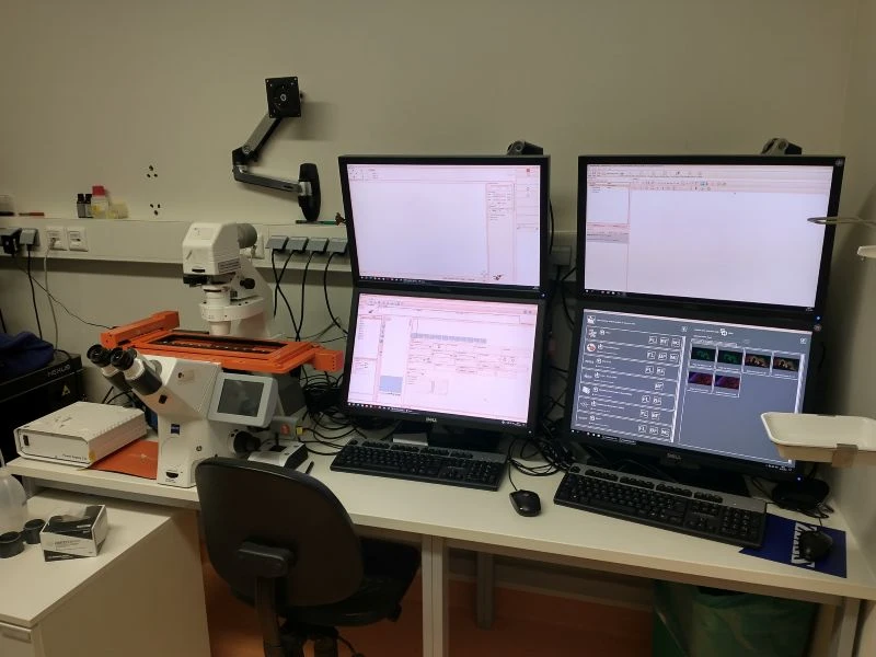 TissueFAXS at the Department of Immunology at Leibniz Institute for Immunotherapy (LIT)
What are your most important reference publications?
- Delacher M, Simon M, Sanderink L, Hotz-Wagenblatt A, Wuttke M, Schambeck K, Schmidleithner L, Bittner S, Pant A, Ritter U, Hehlgans T, Riegel D, Schneider V, Groeber-Becker FK, Eigenberger A, Gebhard C, Strieder N, Fischer A, Rehli M, Hoffmann P, Edinger M, Strowig T, Huehn J, Schmidl C, Werner JM, Prantl L, Brors B, Imbusch CD, Feuerer M. 2021. Single-cell chromatin accessibility landscape identifies tissue repair program in human regulatory T cells. Immunity 54: 702-20 e17
- Schmid M, Dufner B, Durk J, Bedal K, Stricker K, Prokoph LA, Koch C, Wege AK, Zirpel H, van Zandbergen G, Ecker R, Boghiu B, Ritter U. 2015. An Emerging Approach for Parallel Quantification of Intracellular Protozoan Parasites and Host Cell Characterization Using TissueFAXS Cytometry. PLoS One 10: e0139866
- Zimara N, Florian C, Schmid M, Malissen B, Kissenpfennig A, Mannel DN, Edinger M, Hutchinson JA, Hoffmann P, Ritter U. 2014. Langerhans cells promote early germinal center formation in response to Leishmania-derived cutaneous antigens. Eur J Immunol 44: 2955-67