Understanding the Different Types of FISH Evaluations
FISH is a powerful imaging-based technique for detecting DNA and RNA sequences directly in cells and tissues. This post explores the advantages of FISH, its complementary methods like CISH and RNAscope, and how TG solutions streamline automated FISH analysis.
Blog Post
Understanding the Different Types of FISH Evaluations
18 Jan, 2023
Fluorescence in-situ hybridization (FISH) is one of the most powerful tools for detecting genetic aberrations directly within cells and tissues. By using fluorescent probes to target specific DNA or RNA sequences, FISH enables researchers and clinicians to visualize genetic abnormalities with high accuracy and efficiency. Beyond diagnostics, FISH plays a central role in cancer biology, neuroscience, and infectious disease research, and its versatility has driven the development of complementary methods such as CISH and RNAscope. [1].
What are the advantages of performing FISH evaluations?
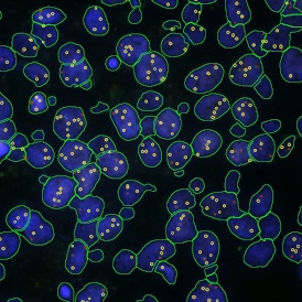
FISH is a sequencing-based method that operates via fluorescent oligonucleotide probes that attach to specific complementary parts of a cell's chromosome and signals its attachment and location via fluorescence imaging [1]. After this data is assessed, scientists can quickly and easily identify gene expression profiles or genetic abnormalities flagged by the probes and analyze what type of abnormality was detected via the FISH evaluations. Because of this capability, FISH possesses high throughput, accuracy, specificity, and is more time efficient in comparison to traditional methods of cell genomics such as karyotype analysis [1].
There are many types of different samples to which FISH can be applied, including tissue samples, cells, microbes, telomeres, to detect DNA damage or even identify the expression of gene patterns in the brain [1]. Furthermore, various types of FISH evaluations can be conducted with advanced tissue cytometry platforms such as TissueFAXS and StrataQuest (TissueGnostics) to extract usable data from these research experiments.
Capabilities of FISH evaluations
Other types of in-situ detection methods were developed to enhance existing FISH evaluations and can further supplement the analysis of complex biomedical research concepts such as cancer and tumor biology. For example, chromogenic in-situ hybridization (CISH) uses enzymatic reactions instead of fluorescence dye (like in FISH) to detect the targets [2]. Another similar method is RNAScope, which is capable of detecting RNA instead of DNA sequences. Precisely in the tumor microenvironment, RNAscope staining can spatially map cells, characterize genetic signatures, classify heterogeneous cell types and circulating tumor cells, and analyze cellular response to cancer drugs [2]. All these processes rely on robust imaging platforms to analyze and quantify the extracted visual data into translatable information for clinical decisions based on FISH evaluations.
TissueGnostics as a leader in developing solutions for FISH evaluations
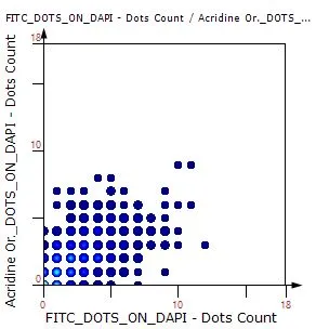
TissueGnostics is a leading life sciences company that provides cutting-edge tissue cytometers as solutions for the acquisition and analysis of different FISH experiments. Experienced in bringing together scientific workflows with innovative imaging and analysis techniques, TissueGnostics allows users to analyze FISH experiments. TissueGnostics offers a variety of automated and streamlined analysis workflows which can be run in their contextual image analysis software StrataQuest:
- IF Dots App – eg for the analysis of FISH experiments (fluorescence mode);
- RNAScope App – eg analysis of dotted structures in brightfield mode;
- Dots analysis combined with other analysis techniques (eg IF Cardio Cell Culture Dots, IHC Small Intestine Dots etc.);
- And many more – check them out here in TissueGnostics App Center.
If you cannot find the perfect solution for your research question please contact TissueGnostics . The scientific experts and technical professionals at TissueGnostics will work together with you to design and deliver the best solutions for your research needs.
References
- Ratan, ZA, Zaman, SB, Mehta, V., Haidere, MF, Runa, NJ, & Akter, N. (2017). Application of Fluorescence In Situ Hybridization (FISH) Technique for the Detection of Genetic Aberration in Medical Science. Cureus. https://doi.org/10.7759/cureus.1325
- Mungenast, F., Fernando, A., Nica, R., Boghiu, B., Lungu, B., Batra, J., & Ecker, RC (2021). Next-Generation Digital Histopathology of the Tumor Microenvironment. Genes, 12(4), 538. https://doi.org/10.3390/genes12040538
- (2022). IF Dots APP: FISH. tissuegnostics.com . https://tissuegnostics.com/en/products/imaging-software/170-tissuefaxs-imaging-software
Are you also interested in a demonstrative case study on FISH image analysis ?
or in How FISH Image Analysis Factors into Next-Gen Digital Pathology ?