Automated Imaging of Metabolic Enzyme Activity in the Skin
Prof. Florian Gruber and Christopher Kremslehner (MedUni Vienna) present a metabolic imaging workflow integrating TissueFAXS i PLUS and StrataQuest to study UV-induced effects on skin (Redox Biology).
Webinar
Automated Imaging of Metabolic Enzyme Activity in the Skin
03 May, 2021
Webinar Transcript
Tissue Cytometry Assisted Metabolic Imaging in Skin
Prof. Florian Gruber & Christopher Kremslehner (Medical University of Vienna / Christian Doppler Laboratories for Skin Aging)
Read assocciated publication
Introduction (Prof. Florian Gruber)
Welcome to our webinar. My colleague Christopher Kremslehner and I will guide you through our projects at the Christian Doppler Laboratories in Skin Aging at the Medical University of Vienna.
As our skin ages, lifestyle factors such as recreational sun exposure and pollution accelerate this process. This aged phenotype is characterized by the appearance of damaged and senescent cells.
At the CD Laboratory, we use a multimodal imaging approach to gain holistic insights into skin aging in human tissue and model systems. Our workflow integrates:
- Mass spectrometry of lipids, metabolites, and molecules (TU Vienna)
- Non-invasive imaging and modeling (BOKU Vienna)
- Automated immunofluorescence and metabolic activity imaging (MedUni Vienna with TissueGnostics technology)
The aim is to correlate these multimodal datasets for a complete picture of cellular damage and senescence within tissue context.
What is Cellular Senescence?
Senescence is a cell-cycle arrest state, distinct from quiescence (G0) or terminal differentiation. It occurs more frequently with age, but is also relevant for embryogenesis, wound healing, and tumor suppression.
Common triggers:
- Persistent DNA damage (e.g. telomere shortening, oncogene activation, chemotherapy)
- Protein and lipid damage (oxidation, cross-linking)
- Reduced nuclear integrity (loss of Lamin B1)
- Mitochondrial dysfunction (metabolic perturbation, proton leaks)
Senescent cells often display metabolic reprogramming, which is harder to detect than DNA or nuclear changes — hence the need for metabolic imaging.
Linking Senescence and Metabolism
Earlier studies showed keratinocytes exposed to UV light reroute metabolism from glycolysis toward the oxidative pentose phosphate pathway (PPP).
- This generates NADPH for antioxidant defense
- Provides nucleotides needed for DNA repair
Our goal was to map these metabolic responses in situ within tissue layers of the epidermis, not just in extracts.
Methods: Skin Models & Tissue Cytometry
We used artificial skin equivalents consisting of epidermal keratinocytes and dermal fibroblasts in a dermal matrix. These models form a stratified epidermis (stratum corneum to basal layer) and can be precisely UV-irradiated.
Damaged, pre-irradiated cells can also be introduced into the model. Tissue sections were then analyzed with:
- Automated IF imaging (TissueFAXS i PLUS)
- StrataQuest image analysis with AI-driven epidermis detection and stratification
- NBT-based colorimetric assays for enzymatic activity (e.g. G6PD)
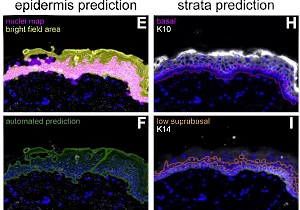
Automated Epidermis Detection (Christopher Kremslehner)
We developed an automated StrataQuest workflow to detect epidermis and sublayers (basal, suprabasal, stratum corneum) using:
- Nuclear density mapping
- Contrast thresholding of brightfield images
- AI classifier-based segmentation (StrataQuest 7)
Validation was performed with keratinocyte markers Keratin 10 (suprabasal) and Keratin 14 (basal), confirming accurate stratum assignment.
Findings
- Baseline metabolism: G6PD activity was higher in basal vs. suprabasal layers, consistent across normal skin and skin models.
- UVB irradiation (150 mJ/cm²):
- Significant increase in G6PD activity across all strata within 1–10 minutes post-UV exposure.
- High G6PD activity co-localized with γH2AX, a marker of DNA damage.
- Suggests metabolic rerouting supports antioxidant defense and DNA repair.
- Fate of damaged keratinocytes:
- Pre-irradiated cells were excluded from basal/suprabasal layers in skin models.
- After one week, these cells showed reduced enzymatic activity compared to neighbors.
- Indicates removal via differentiation and exhaustion of metabolic activity.
Outlook
With StrataQuest 7, AI-based epidermis detection further streamlines workflows. Future work will integrate automated classification and combine it with stratified metabolic analysis for faster, more reproducible results.
Conclusion
- Cellular senescence is closely tied to metabolic reprogramming in skin.
- Metabolic imaging + Tissue Cytometry (TissueFAXS + StrataQuest) allows in-situ mapping of these changes at single-cell resolution.
- This approach provides insights into UV-induced skin damage, aging, and repair mechanisms.
Reference:
Kremslehner C., Gruber F. et. al Imaging of metabolic activity adaptations to UV stress, drugs and differentiation at cellular resolution in skin and skin equivalents – Implications for oxidative UV damage Redox Biology, Volume 37, 2020, https://doi.org/10.1016/j.redox.2020.101583