Exploring metabolic reprogramming via brightfield image analysis
Cells are inherently responsive, shaped by internal priorities and external stimuli. One of the most compelling expressions of this adaptability is metabolic reprogramming, a cellular strategy that involves shifting energy production and biosynthetic activity to support survival, repair, immune activation, or rapid proliferation.
Blog Post
Exploring metabolic reprogramming via brightfield image analysis
24 Oct, 2025
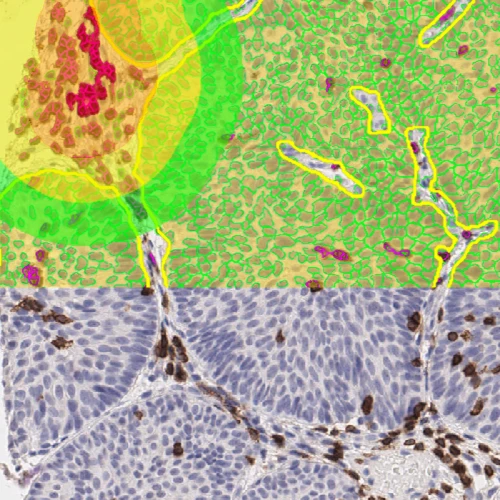
These metabolic shifts influence cellular structure, alter tissue organization, and leave morphological footprints that can be directly observed under a microscope. These changes are usually best visualized using various histological stains, such as hematoxylin and eosin (H&E) staining, or marker-specific, such as immunohistochemical (IHC) staining using specific antibodies.
When brightfield imaging is used in conjunction with the precision of image analysis, researchers can uncover both the form and function of metabolism changes in situ. This integration, defined as brightfield image analysis, reveals not just what metabolic changes are occurring, but exactly where and how they unfold within the native tissue environment.
Interpreting Metabolic Change Through Structural Cues
As cells adjust their energy pathways, they may grow in size, change shape, or express different markers. These morphological shifts often provide the earliest visual evidence of metabolic adaptation and can be captured with standard light microscopy.
Brightfield image analysis transfers these structural transformations in actual numbers. Image analysis of images acquired from IHC-stained tissues also enhances data reliability through quantifying subtle expression differences that might be overlooked.
Together, these methods offer a view of metabolism that connects physical form with underlying biochemical activity through image analysis.
Histological Stains as Metabolic Indicators
Histological stains offer a layered perspective on cellular metabolism, making invisible biochemical shifts visible within the structural fabric of tissues. Lipid accumulation, a hallmark of altered fatty acid metabolism, is revealed through stains like Oil Red O and Sudan Black. PAS staining exposes glycogen distribution, uncovering how cells store or mobilize glucose. Meanwhile, H&E staining provides broader context, revealing tissue-level disruptions like necrosis and inflammation that signal metabolic imbalance.
Brightfield image analysis captures the spatial distribution of these stains, mapping metabolic activity across tissue regions. This spatial understanding becomes more powerful with numerical data, which quantifies staining intensity, localization, and variability.
Illuminating Molecular Drivers of Metabolism
To fully characterize metabolic reprogramming, it's essential to move beyond morphology and directly observe the molecules at its core. IHC allows the visualization of enzymes and transporters including GLUT1, LDHA, CPT1A, and FASN. These markers play defining roles in metabolic pathways including glycolysis, lipogenesis, and fatty acid oxidation.
The combined analysis of IHC data and structural context allows researchers to assess expression levels within distinct tissue regions and compare metabolic profiles across experimental conditions.
Making Metabolism Measurable with TissueGnostics
Understanding metabolic reprogramming requires tools that illuminate the visible and quantify the invisible. Brightfield image analysis transforms ordinary tissue sections into dynamic metabolic maps. This dual capability enables researchers to uncover how metabolic pathways manifest structurally within biological tissues and how they can be quantified in spatial context.
TissueGnostics supports this integrated approach with platforms like the TissueFAXS PLUS. This system delivers high-resolution brightfield and fluorescence whole-slide-imaging for tissue sections on slides, smears, cytospins, and tissue microarrays. Paired with software such as StrataQuest, it enables automated image analysis and supports everything from focused tissue exploration to high-throughput metabolic screening.
As imaging and analysis converge, metabolism becomes a clearly defined spatial and functional element of tissue biology, offering clarity and momentum for scientific discovery.