
IHC Adipocytes+
The IHC Adipocytes+ App detects adipocytes and cellular aggregates between them. Outputs include number and area measurements for adipocytes and aggregates.
single-cell analysis
adipocytes, fat tissue, fat cells, immunohistochemistry, immune cell detection
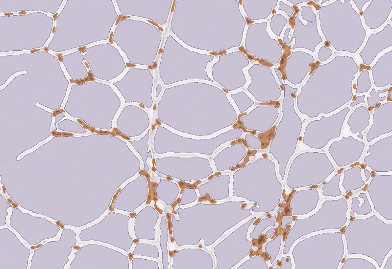
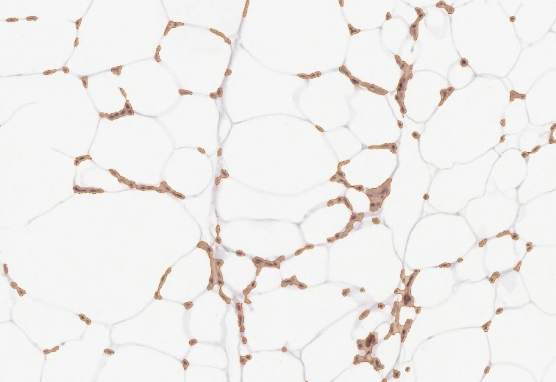
The IHC Adipocytes+ App identifies adipocytes and cellular aggregates inbetween the adipocytes. Small rips in adipocyte membranes are mended automatically and cell membrane artefacts in adipocyte lumina are automatically eliminated. The App outputs number and area measurements for all detected adipocytes as well as number and area of cellular aggregates.
Original Image
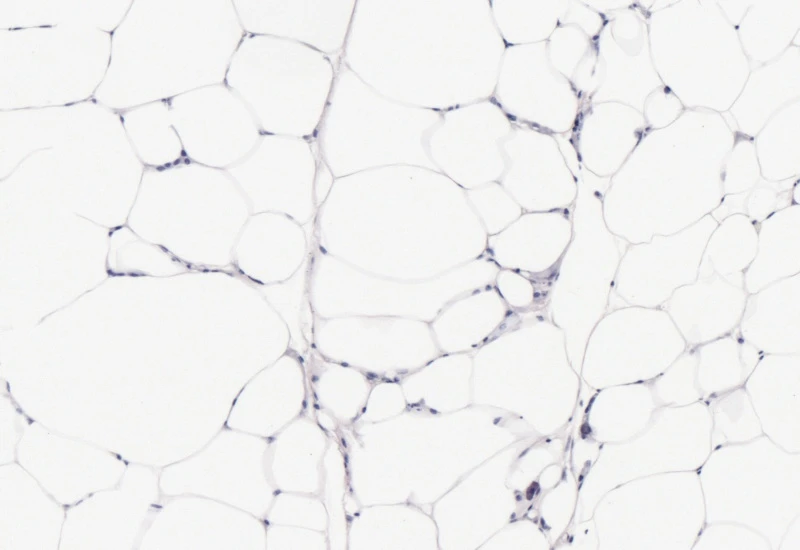
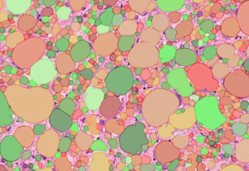
Adipocyte detection
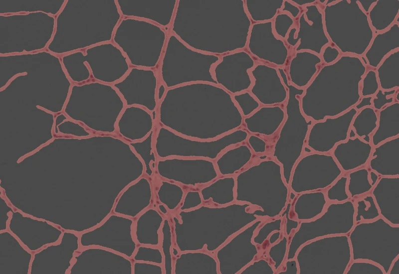
Cluster detection
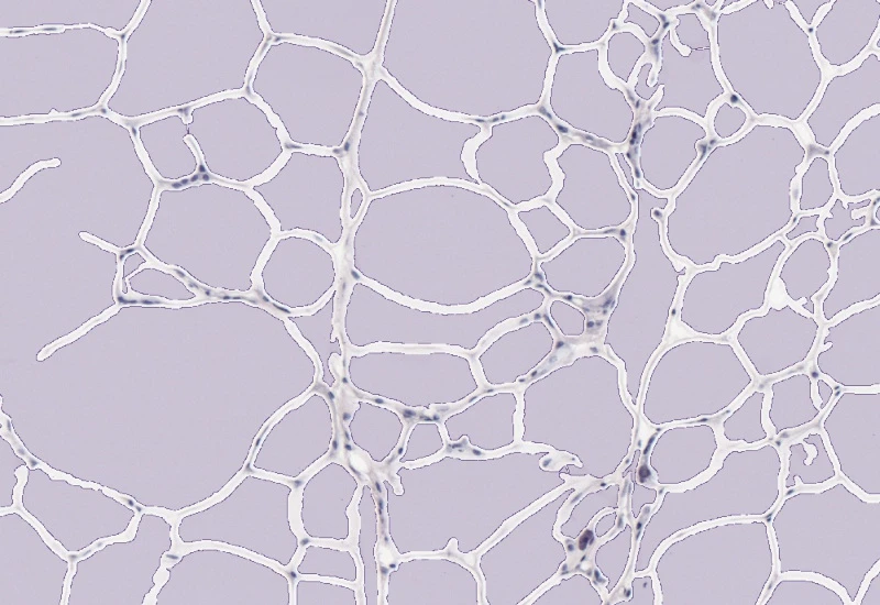
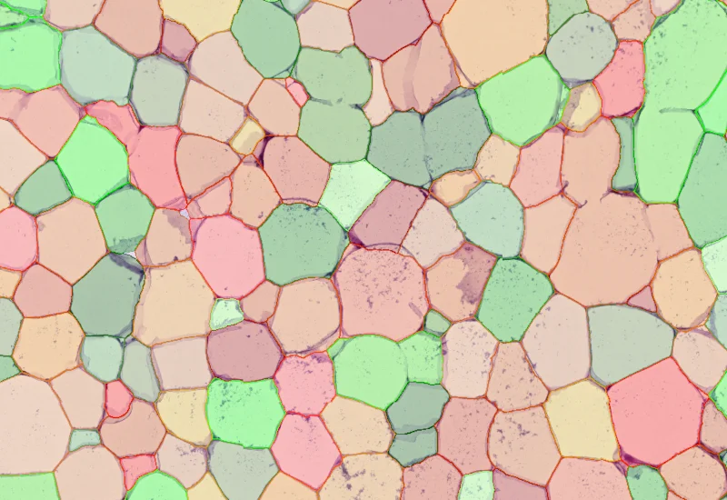
Combined detection
single-cell analysis
Blog Post
23 Jul, 2026
How Histology Slide Scanners are Used to Study Osteoarthritis
We support the following file formats:
- TissueFAXS (aqproj)
- StrataFAXS II (vmic)
- PreciPoint (vmic, gtif)
- Generic BigTIFF Import
- Support for multipage BigTIFF files
- OME-TIFF
- JPEG, PNG, BMP, TIFF
- Zeiss (czi)
- Hamamatsu NanoZoomer (ndpi)
- Aperio (svs)
- Leica (scn)
- 3D HISTECH Pannoramic
- Mirax (mrxs)
- Olympus (vsi)
- More slide scanners to be added!
Related Apps
Lipid Droplets
The Lipid Droplets App quantifies lipid droplets in H&E-stained tissues, correcting membrane artifacts and providing counts and area measurements.
liver, lipid droplets, H&E, brightfield


Custom App development
Perfectly tailored image analysis solutions for your research.
You have a specific research question that needs to be answered? We offer custom development of image analysis pipelines for specific tasks, be it detection of cellular phenotypes or quantification of tissue structures. After discussing your goals with one of our experts, you will get a ready-to-use App and be a step closer to an impactful publication.