
IF Cellular microenvironment
Determine phenotypes of specific IF-stained cell populations, analyze spatial relationships to neighboring cells and metastructures (e.g. vessels, tumors), and perform proximity and infiltration analyses.
metastructures
single-cell analysis
tumor microenvironment
multiplex IF
immunophenotyping
phenotyping, phenotype interactions, proximity map, spatial landscape, immune cells, tumor, colon cancer, TMA, Foxp3, CD4, CK, PD-1, T regulatory cells, fluorescence
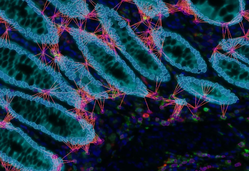
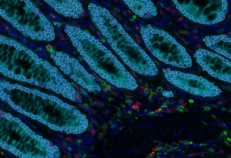
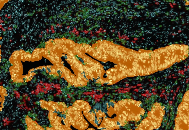
The IF Cellular Microenviroment App allows to determine the cellular phenotype of specific IF stained cell populations and establishes their spatial relationship between each other, their neighbour cells/cell populations as well as the one with metastructures (e.g. blood vessels, tumors) in their vicinity. It is especially suited for proximity and infiltration analyses.
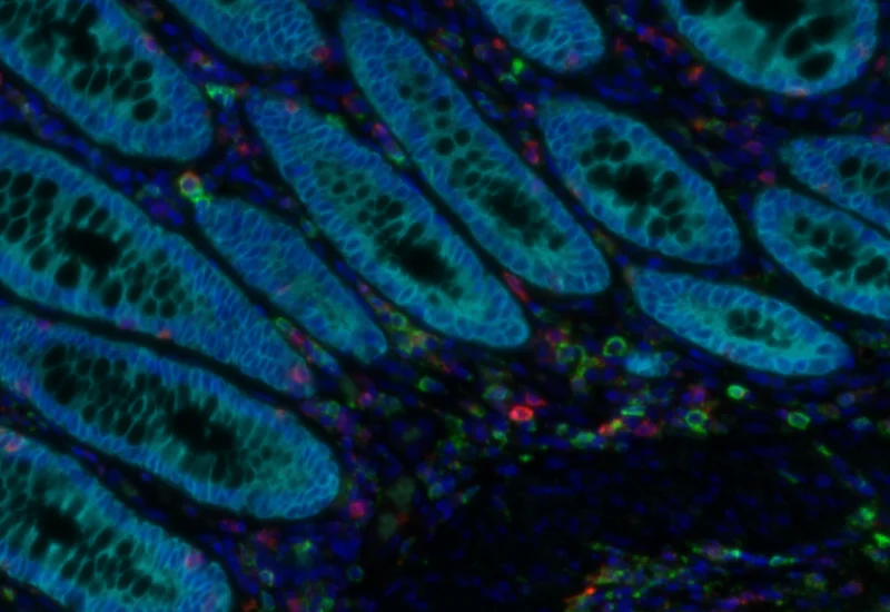
Original image
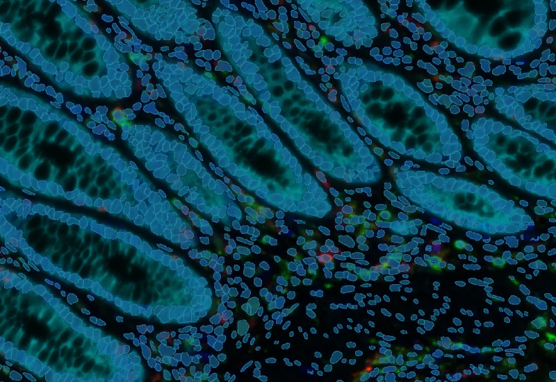
Nuclei segmentation
Epithelium detection
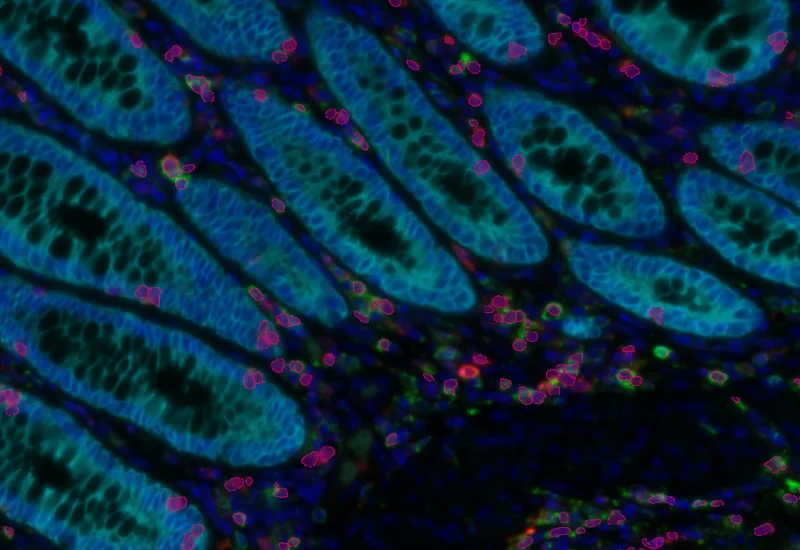
Phenotype detection
Phenotype interaction
immunophenotyping
Application Note
06 Jul, 2026
Mapping spatial cell-to-cell interactions in tertiary lymphoid structures with spatial proteomics
multiplex IF

White Paper
01 Oct, 2026
A New Neutrophil Subset Suppresses Immunity Response in CRCLM
tumor microenvironment
White Paper
01 Oct, 2026
A New Neutrophil Subset Suppresses Immunity Response in CRCLM
single-cell analysis

Blog Post
23 Jul, 2026
How Histology Slide Scanners are Used to Study Osteoarthritis
metastructures
Blog Post
17 May, 2023
An Intro to Deep Learning in Biomedical Imaging
We support the following file formats:
- TissueFAXS (aqproj)
- StrataFAXS II (vmic)
- PreciPoint (vmic, gtif)
- Generic BigTIFF Import
- Support for multipage BigTIFF files
- OME-TIFF
- JPEG, PNG, BMP, TIFF
- Zeiss (czi)
- Hamamatsu NanoZoomer (ndpi)
- Aperio (svs)
- Leica (scn)
- 3D HISTECH Pannoramic
- Mirax (mrxs)
- Olympus (vsi)
- More slide scanners to be added!
Related Apps
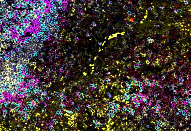
IF Cellular Contact
Determine cellular phenotypes in IF-stained samples, analyze direct cell–cell contacts, and quantify marker intensity, morphometric parameters, and number/% of contacting phenotypes.
cell contacts, spatial interactions, phenotype interactions, B cells, T cells, lung, lymph node, COVID-19, SARS-Cov-2
IF Immune status in situ
Characterize immune cell phenotypes relative to detected metastructures (e.g. tumors, glands), define distance ranges, measure cell-to-boundary distances inside/outside, and export up to 20 intensity, statistic, and morphometric parameters per cell compartment.
colon cancer, cytotoxic t cells, tumor microenvironment, PD1, CD8, spatial analysis, fluorescence

IHC Immune Status in Situ
Segment tissue into tumor, stroma, and lymphocyte clusters using an AI classifier, detect hematoxylin-stained nuclei and immune phenotypes (e.g., CD45, CD3, CD20), and quantify spatial immune distribution.
bladder cancer, CD45, immune cells, tumor immune microenvironment, tertiary lymphoid structures, spatial analysis

Custom App development
Perfectly tailored image analysis solutions for your research.
You have a specific research question that needs to be answered? We offer custom development of image analysis pipelines for specific tasks, be it detection of cellular phenotypes or quantification of tissue structures. After discussing your goals with one of our experts, you will get a ready-to-use App and be a step closer to an impactful publication.


