
IF Artificial Skin
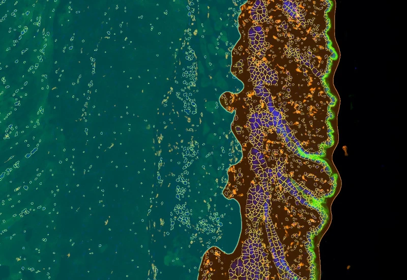
Stratify skin equivalents into dermis and epidermis, segment the stratum corneum, high/low suprabasal and basal layers, and quantify staining intensity, area, nuclei, and number/% of marker-positive cells for each layer.
metastructures
single-cell analysis
dermatology
dermatology, epidermis, dermis, skin, aging, oxidative UV damage, artificial skin
The IF Artificial Skin App stratifies skin equivalents into differnet layers including dermis and epidermis and identifies nuclei based on appropriate staining. Further is subclassifies epidermis into stratum corneum, high suprabasal, low suprabasal, basal. The App outputs, area (µm2) of epidemis and dermis, area and mean staining intensity for each sublayer of the epidermis, total number of nuclei for each layer/sublayer and number/% of marker positive cells for each layser and sublayer.
Kremslehner C, Miller A, Nica R, et al. Imaging of metabolic activity adaptations to UV stress, drugs and differentiation at cellular resolution in skin and skin equivalents - Implications for oxidative UV damage. Redox Biol. 2020;37:101583. doi:10.1016/j.redox.2020.101583
Image courtesy of Prof. Florian Gruber
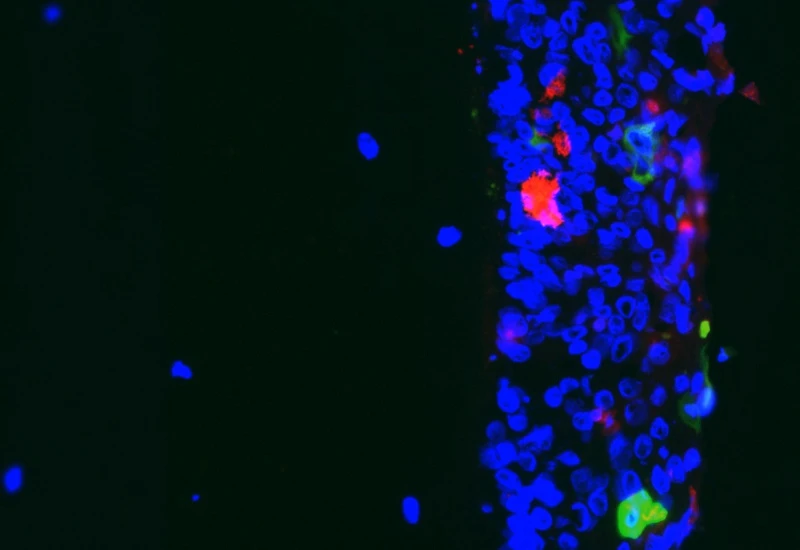
Original image
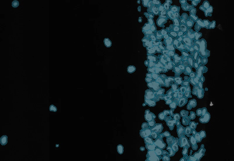
Detection of nuclei

Detection of dermis and epidermis
Detection of epidermis sublayers
dermatology
Webinar
21 Oct, 2025
Multimodal Imaging of Cellular Senescence: Tissue Cytometry and Beyond
single-cell analysis

Blog Post
23 Jul, 2026
How Histology Slide Scanners are Used to Study Osteoarthritis
metastructures

Blog Post
17 May, 2023
An Intro to Deep Learning in Biomedical Imaging
We support the following file formats:
- TissueFAXS (aqproj)
- StrataFAXS II (vmic)
- PreciPoint (vmic, gtif)
- Generic BigTIFF Import
- Support for multipage BigTIFF files
- OME-TIFF
- JPEG, PNG, BMP, TIFF
- Zeiss (czi)
- Hamamatsu NanoZoomer (ndpi)
- Aperio (svs)
- Leica (scn)
- 3D HISTECH Pannoramic
- Mirax (mrxs)
- Olympus (vsi)
- More slide scanners to be added!

Custom App development
Perfectly tailored image analysis solutions for your research.
You have a specific research question that needs to be answered? We offer custom development of image analysis pipelines for specific tasks, be it detection of cellular phenotypes or quantification of tissue structures. After discussing your goals with one of our experts, you will get a ready-to-use App and be a step closer to an impactful publication.