HE Alveoli Hamster
Detect nuclei and alveoli in HE-stained lung sections, classify alveoli into user-defined size ranges, and quantify tissue area, total alveoli area, and number/% per size class. Adaptable to other species.
brightfield
metastructures
hamster, rodents, H&E, lung, aveoli, pneumology
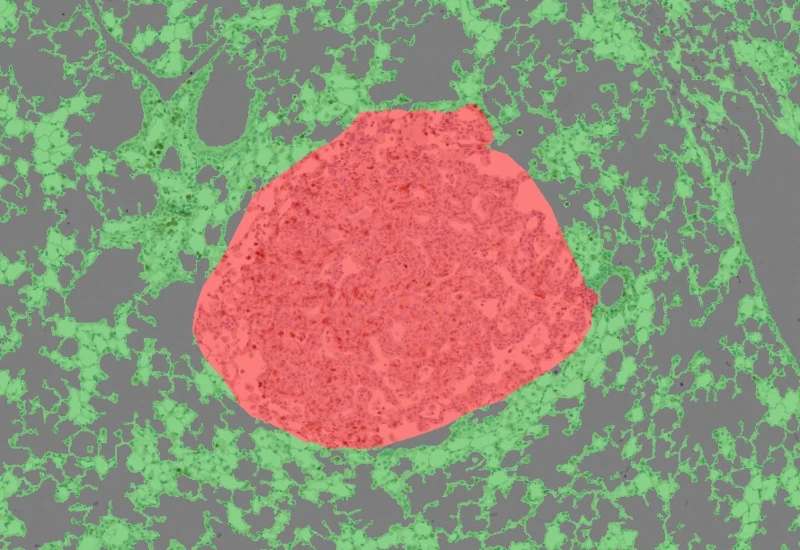
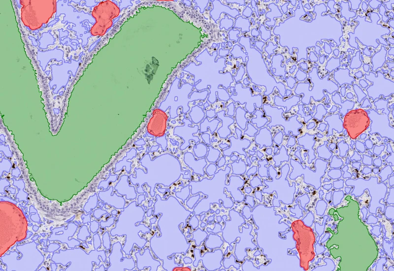
The HE Aveoli Hamster App provides detection of nuclei, aveoli, and the categorization of aveoli in user defined size classes (e.g. 0-1000µm2, 1000-8000µm2, 8000-15000µm2) in HE stained tissue sections. It outputs total tissue area (µm2), total aveoli area (µm2), number and % of aveoli for user-defined size classes. This App can be easily adapted to tissues of other species such as mouse or rat.
Image courtesy of Marc Scott, TRI, Queensland University
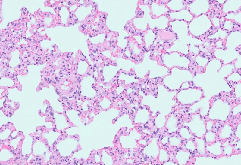
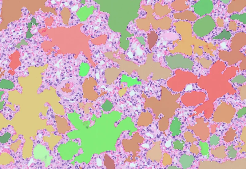
Original Image
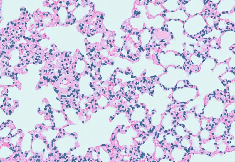
Detection of nuclei
Detection of alveoli
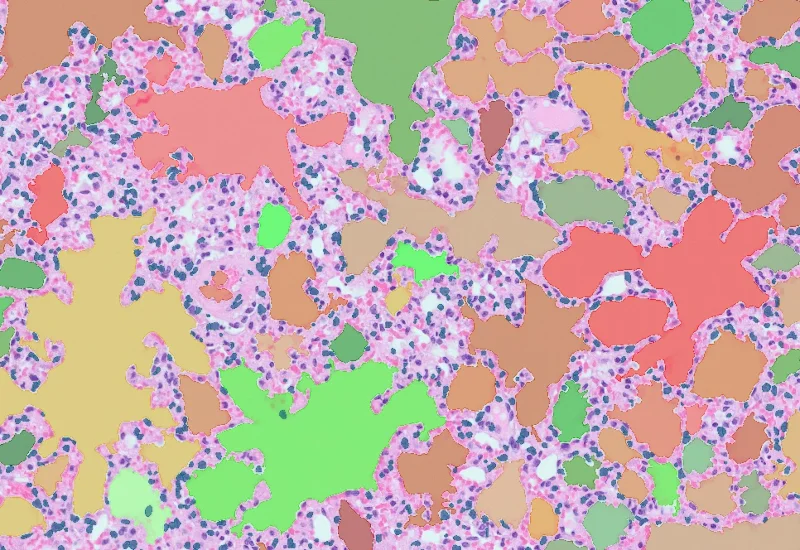
Combined detection
metastructures

Blog Post
17 May, 2023
An Intro to Deep Learning in Biomedical Imaging
We support the following file formats:
- TissueFAXS (aqproj)
- StrataFAXS II (vmic)
- PreciPoint (vmic, gtif)
- Generic BigTIFF Import
- Support for multipage BigTIFF files
- OME-TIFF
- JPEG, PNG, BMP, TIFF
- Zeiss (czi)
- Hamamatsu NanoZoomer (ndpi)
- Aperio (svs)
- Leica (scn)
- 3D HISTECH Pannoramic
- Mirax (mrxs)
- Olympus (vsi)
- More slide scanners to be added!
Related Apps
IHC Lung Cancer Mouse
Segment murine lung sections into tumor and non-cancerous tissue using a machine learning classifier, detect hematoxylin-stained nuclei, and quantify tumor area and marker-positive cellular phenotypes.
immunohistochemistry, lung cancer, tumor microenvironment, mouse, lung
Pulmo
Segment nuclei and lung metastructures (total tissue, bronchioles, vessels, alveoli), detect cellular phenotypes within each component, and measure up to 20 morphometric parameters per metastructure and nucleus.
lung cancer, lung anatomy, bronchioles, blood vessels, alveoles, immunohistochemistry, neutrophiles, brightfield

Custom App development
Perfectly tailored image analysis solutions for your research.
You have a specific research question that needs to be answered? We offer custom development of image analysis pipelines for specific tasks, be it detection of cellular phenotypes or quantification of tissue structures. After discussing your goals with one of our experts, you will get a ready-to-use App and be a step closer to an impactful publication.