Dynamic Content Image

Bone von kossa

Detect mineralized bone in von Kossa–stained sections and measure TV, BV, BS, BV/TV, BS/BV, Tb.N, Tb.Th, and Tb.Sp.

mineralized bone, bone research, von kossa, osteoporosis, brightfield

Dynamic Content Image

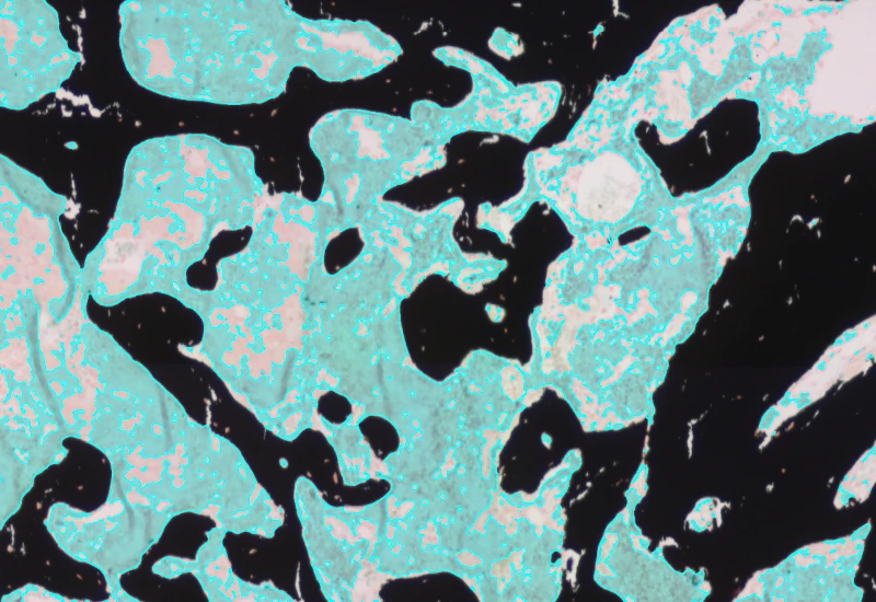
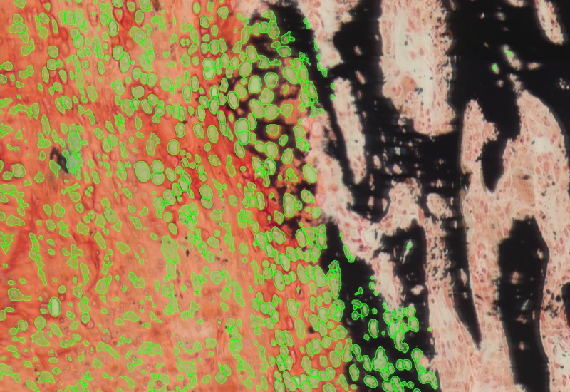
The Bone Tissue Analysis Von Kossa APP allows to detect mineralized bone tissue based on ‘von kossa’ stained bone tissue sections. The APP provides parameters such as TV, BV, BS, BV/TV, BS/BV, Tb.N, tb.Th, Tb.Sp.
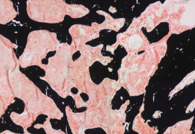
Dynamic Content Image

Original image

Dynamic Content Image

Mineralized bone detection

Dynamic Content Image

Bone marrow detection

Dynamic Content Image

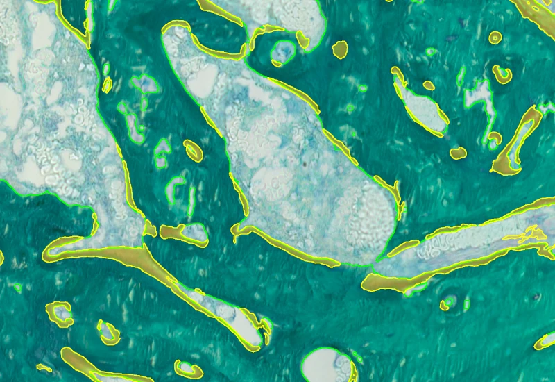
Bone surface detection

Dynamic Content Image

White Paper

17 Jul, 2024

Algorithmic automation of bone research with StrataQuest

Dynamic Content Image

Blog Post

17 May, 2023

An Intro to Deep Learning in Biomedical Imaging

We support the following file formats:

  • TissueFAXS (aqproj)
  • StrataFAXS II (vmic)
  • PreciPoint (vmic, gtif)
  • Generic BigTIFF Import
  • Support for multipage BigTIFF files
  • OME-TIFF
  • JPEG, PNG, BMP, TIFF
  • Zeiss (czi)

  • Hamamatsu NanoZoomer (ndpi)
  • Aperio (svs)
  • Leica (scn)
  • 3D HISTECH Pannoramic
  • Mirax (mrxs)
  • Olympus (vsi)
  • More slide scanners to be added!

Related Apps

Dynamic Content Image

Bone Goldner

Detect mineralized bone and osteoid in Goldner-stained sections and measure BV, TV, OV, OV/BV, OV/TV, OS, BS, mean osteoid width, and mean osteoid thickness.

metastructures

bone research

goldner staining, mineralized bone, osteoid, BV, TV, OV, OS, BS, brightfield

Dynamic Content Image

Bone Mineralization

Separate Safranin O–stained bone into cartilage, mineralized cartilage, bone marrow, and mineralized bone, and measure TV, BV, MCV, CV, and BM.

metastructures

bone research

Safranin O, bone mineralization, bone marrow, cartilage, TV, BV, MCV, CV, BM, brightfield

Dynamic Content Image

IF Cell Culture - Osteoclast

Segment nuclei, identify multinucleated osteoclasts, and quantify cell number, nuclei per osteoclast, osteoclast area, and marker intensity within osteoclasts.

single-cell analysis

bone research

cell culture

bone research, osteoclast, multinucleated cells, bone adsoprtion

Image

Custom App development

Perfectly tailored image analysis solutions for your research.

You have a specific research question that needs to be answered? We offer custom development of image analysis pipelines for specific tasks, be it detection of cellular phenotypes or quantification of tissue structures. After discussing your goals with one of our experts, you will get a ready-to-use App and be a step closer to an impactful publication.

LogoOrangewhite2025

TissueGnostics GmbH
Taborstraße 10/2/8
1020 Vienna, Austria
+43 1 216 11 90
This email address is being protected from spambots. You need JavaScript enabled to view it.

Contact us

About Us

TissueGnostics provides advanced solutions for whole-slide imaging and image analysis in biological and clinical research. Our products help researchers to scan and analyze complex tissue samples, enabling more detailed insights into tissue structure, cellular interactions, and spatial cell landscape.

Keep in Touch

Be the first to hear about the latest scientific updates.

We use cookies

We use cookies on our website. Some of them are essential for the operation of the site, while others help us to improve this site and the user experience (tracking cookies).
You can decide for yourself whether you want to allow cookies or not. Please note that if you reject them, you may not be able to use all the functionalities of the site.