IBEX and StrataQuest image analysis compatibility
May, 2022
One of the challenges of imaging tissue sections is dealing with their diverse composition. The human body contains hundreds of different cell phenotypes, with many more to be discovered. There is a huge need for multiplexing imaging techniques that can recover imaging information on cell-cell interactions with sufficient resolution to identify various unique phenotypes and is compatible with the diversity of cell species.
IBEX Protocol
The recently developed Iterative Bleaching Extends multi-plexity (IBEX) imaging method offers a powerful approach to the multiplex imaging of diverse tissues, including human organs.1,2 IBEX is a relatively low-cost protocol that takes 2-5 days to complete for biologists with just basic laboratory skills and is designed to recover ultra-high content information for quantitative and qualitative analysis.
At the core of the IBEX protocol is a multiplex antibody-based imaging method that uses commercially available reagents and equipment for the spatial characterization of complex phenotypes in tissue. While manual processing of IBEX images is possible, the method can also be used with automated content imaging and sample processing.
One of the key advantages of the IBEX protocol is the flexibility and diversity of the method. It is compatible with different imaging platforms and 250 commercially available antibodies, and 16 fluorophores. Protocols recommending antibody-fluorophore combinations and tissue preparation steps also exist to encourage widespread adoption of the method and minimize the amount of characterization and preparatory work that needs to be performed by individual laboratories.
The short immunolabelling and bleaching times are another benefit of IBEX imaging. The ability to stain and image many biomarkers in a single tissue sample is crucial to exploring the full complexity of most tissue samples.
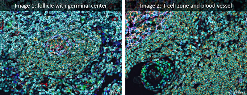
Analyzed lymph node images were acquired using the iterative bleaching extends multiplexity (IBEX) method (PNAS, Nature Protocols), an open source multiplexed antibody-based imaging method (Nature Methods). StrataQuest analysis was performed on a lymph node dataset shared publicly by the authors (Accompanying Zenodo).
PNAS: https://www.pnas.org/doi/10.1073/pnas.2018488117Nature Protocols: https://www.nature.com/articles/s41596-021-00644-9Nature Methods: https://www.nature.com/articles/s41592-021-01316-yAccompanying Zenodo: https://zenodo.org/record/5244551#.YnVAZ-jMIuU
StrataQuest
One way to enhance the efficiency, throughput potential and in-depth analysis of the images from IBEX protocols is to make use of automated image analysis.
StrataQuest is TissueGnostics’ world-leading analysis software for brightfield and fluorescence image analysis.3
StrataQuest has been designed with simplifying and accelerating workflows in mind. To use StrataQuest to analyze the output of an IBEX protocol, that workflow might first start with image alignment. IBEX procedures can generate either 3D image stacks or 2D single-z slices.
The alignment step is often performed using the intensity of the images or in multi-cycle IBEX experiments, by analyzing the image to find the presence of a repeated marker to register all images to a fixed position. This marker can be a number of features – either a particular cell organelle or structural feature.
Normalization of the images is performed using a similarity metric on the aligned images, providing a robust registration of a large volume of data in three dimensions of data. The protocol and its implementation with StrataQuest can process over 160 GB of data with over 20 imaging cycles in just a few minutes.
Simple Interfacing
Once images have been processed, these can be analyzed to explore the tissue samples. Within StrataQuest, it is possible to build workflows that incorporate the necessary image processing for handling the output of IBEX experiments and can be used to automate subsequent data analysis workflows. The analysis part could include for instance in-depth tissue classification, immunophenotyping and spatial analysis.
The StrataQuest predefined workflow can be set to export any results in the end of the analysis in a suitable format. StrataQuest 7.1 has many inbuilt analysis engines, including a machine learning-based tissue classifier or a deep learning based nuclei segmentor, and thereby makes image analysis more accessible for researchers with no background in image analysis.
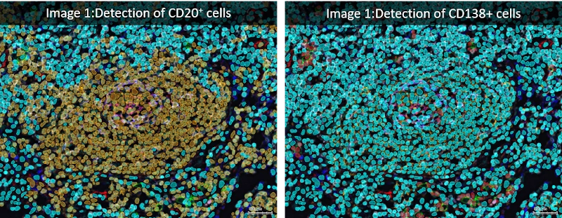
StrataQuest provides the analysis capabilities for cell, tissue and structure recognition as well as spatial analysis and works particularly well with IBEX processed samples. The range of tissue types that can be given distinct stains in a single image (with the creation of up to 29 parameter datasets with automated IBEX) means the automated analysis performed in StrataQuest can help accelerate workflows and ensure all potential interactions are captured.
IBEX procedures are particularly powerful for the visualization of tumor-immune interactions that are crucial for understanding the underlying biochemical mechanisms involved in tumor growth and the development of new therapies to target particular types of metastatic cells. A large number of image cycles with IBEX does not lead to cellular degradation and so it is possible to express large numbers of antibodies when many distinct cell types are present in complex human tissues.
TissueGnostics
StrataQuest can be either a standalone analysis program or integrated with imaging equipment to perform automated experiment control. This can include processes such as auto sampling to identify regions of interest for further imaging using the StrataQuest App to identify ‘meaningful objects’. This makes StrataQuest an obvious software choice to support either automated or manual IBEX workflows to extract the maximum information from IBEX images in the minimum time.
References and Further Reading
- Radtke, A. J., Chu, C. J., Yaniv, Z., Yao, L., Marr, J., Beuschel, R. T., Ichise, H., Gola, A., Kabat, J., Lowekamp, B., Speranza, E., Croteau, J., Thakur, N., Jonigk, D., Davis, J., Hernandez, J. M., & Germain, R. N. (2021). IBEX: An open and extensible method for high content multiplex imaging of diverse tissues.
- Radtke, A. J., Kandov, E., Lowekamp, B., Speranza, E., Chu, C. J., Gola, A., Thakur, N., Shih, R., Yao, L., Yaniv, Z. R., Beuschel, R. T., Kabat, J., Croteau, J., Davis, J., Hernandez, J. M., & Germain, R. N. (2020). IBEX: A versatile multiplex optical imaging approach for deep phenotyping and spatial analysis of cells in complex tissues. Proceedings of the National Academy of Sciences of the United States of America, 117(52), 33455–33465. https://doi.org/10.1073/PNAS.2018488117
- TissueGnostics (2022) StrataQuest, https://tissuegnostics.com/products/contextual-image-analysis/strataquest, accessed March 2022.