
Tumor-Derived Exosomal TAGLN2 Promotes Metastasis in Gastric Cancer
Recent research suggests that tumor cells communicate with surrounding tissues long before metastatic lesions become established, creating conditions that favor tumor dissemination and growth. One of important mediators of this communication are extracellular vesicles, particularly exosomes.
tumor microenvironment
oncology
multiplex IF
White Paper
Tumor-Derived Exosomal TAGLN2 Promotes Metastasis in Gastric Cancer
18 Aug, 2026
Gastric cancer (GC) ranks among the most prevalent cancers globally. While primary tumors can often be managed through surgery and systemic therapies, distant metastasis is responsible for the majority of gastric cancer-related deaths.
Recent research suggests that tumor cells communicate with surrounding tissues long before metastatic lesions become established, creating conditions that favor tumor dissemination and growth. One of important mediators of this communication are extracellular vesicles, particularly exosomes (1).
Tumor-derived exosomes (TEX) are nanoscale vesicles that transport proteins and nucleic acids and remodel the tumor microenvironment, influencing immune responses and altering vascular function to support cancer progression (2). Recent research has identified an important new mechanism by which gastric cancer-derived exosomes promote metastasis through the delivery of Transgelin-2 (TAGLN2), a cytoskeletal regulatory protein (3).
Exosomal TAGLN2: A New Driver of Metastatic Progression
Yu et al. showed that TAGLN2 is highly expressed in gastric cancer cells and tumor-associated endothelium. Importantly, elevated endothelial TAGLN2 expression was associated with lymph node metastasis and poorer patient outcomes (3).
The study revealed that exosomes released by gastric cancer cells transport TAGLN2 directly to endothelial cells, where it triggers increased angiogenesis, disruption of endothelial junctions, and enhanced vascular permeability. This ultimately accelerates metastatic spread, which facilitates tumor cell intravasation and dissemination to distant organs (3).
The researchers identified the mechanism of a novel signaling pathway involving Neuropilin-1 (NRP1), Semaphorin-4D (SEMA4D) and yes-associated protein (YAP). Exosomal TAGLN2 induces expression of NRP1 and SEMA4D in endothelial cells.
NRP1 and SEMA4D then form a complex that activates YAP, driving endothelial remodeling and vascular destabilization (3). These findings improve our understanding of gastric cancer biology and highlight potential therapeutic targets and biomarkers that could be exploited for future patient management.
Why Multiplex Immunofluorescence Remains Essential
As cancer biology research becomes increasingly complex, researchers require technologies capable of visualizing multiple biomarkers simultaneously within intact tissue architecture. While genomic and transcriptomic approaches provide valuable molecular information, they often lack the spatial context needed to understand cellular interactions within the tumor microenvironment (4).
Multiplex immunofluorescence (mIF) addresses this challenge by detecting multiple protein markers in a single tissue section, enabling identification of cell populations and their spatial relationships between tumor, stromal, and immune cells (5).
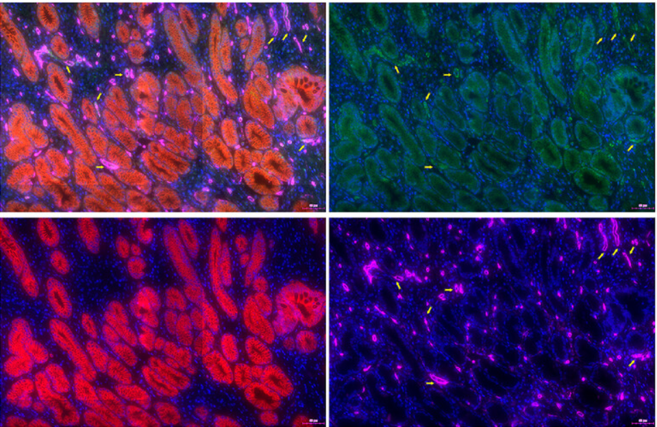
In the TAGLN2 study, investigators used multiplex immunofluorescence (mIF) to analyze tissue samples from 90 patients with gastric cancer together with matched normal gastric tissues. Using a four‑marker panel (TAGLN2, CD34, CK, DAPI) in 90 paired gastric samples, researchers localized TAGLN2 to tumor and vascular compartments, supporting its role in endothelial reprogramming and metastasis (3).
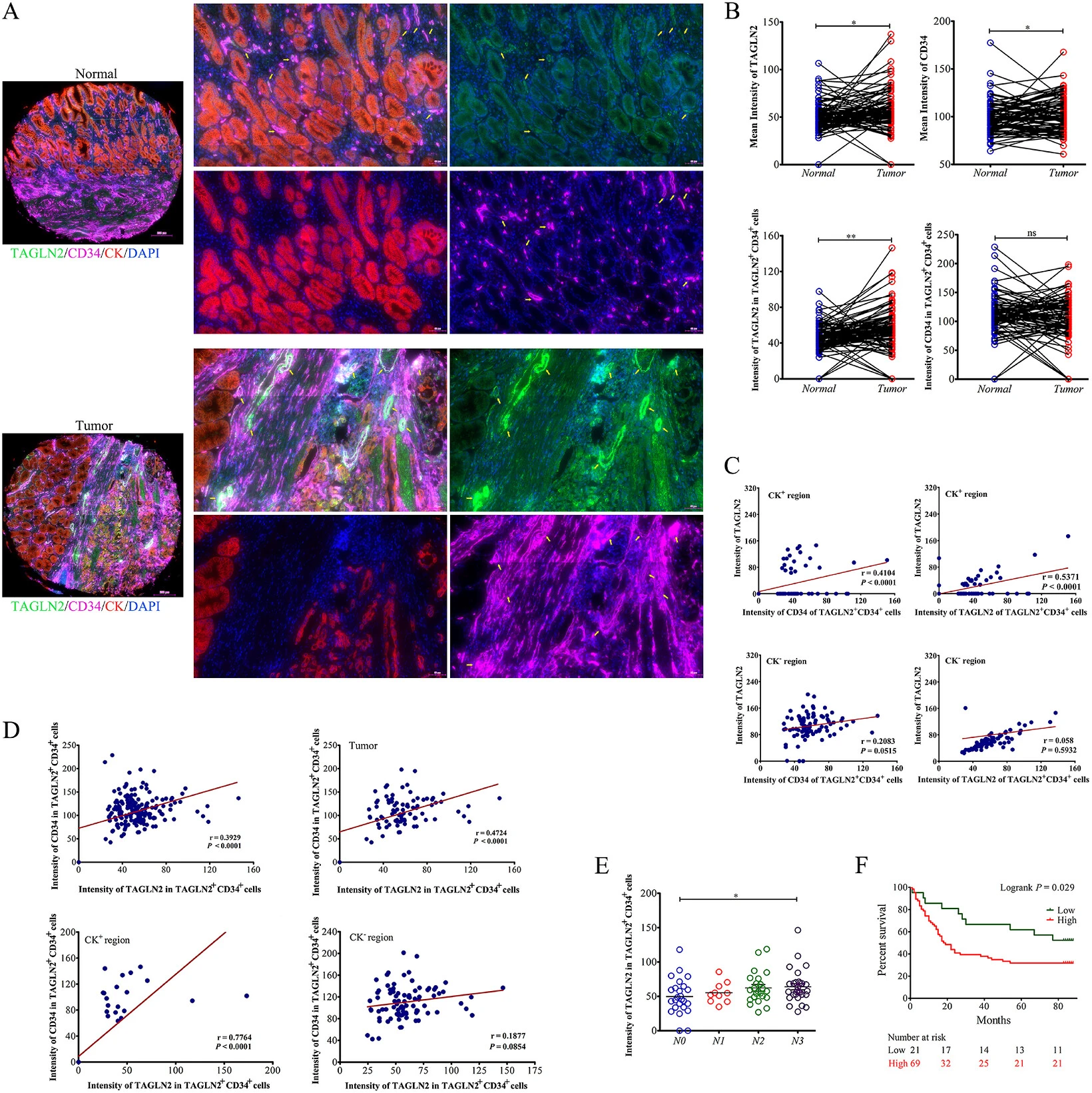
Figure 1. Aberrantly increased expression of TAGLN2 in GC microenvironment. Adapted from: S. Yu, J. Zhuo, X. Hong, et al. “Tumor-Derived Exosomal TAGLN2 Promotes Metastasis by Inducing Vascular Permeability and Angiogenesis via the NRP1/SEMA4D/YAP Axis.” Advanced Science 13, no. 29 (2026): e21962.
From Imaging to Quantitative Insights
Generating high-quality multiplex images is only one part of a successful spatial biology workflow. Equally important is the ability to convert image data into practical quantitative information.
For this study, stained tissue sections were imaged using the TissueFAXS Spectra slide scanner and analyzed using StrataQuest image analysis software. Together, these technologies provide a comprehensive workflow from image acquisition through to advanced quantitative analysis.
TissueFAXS Spectra can detect eight fluorescent markers simultaneously, with plans to expand to 10. Advanced spectral unmixing algorithms separate overlapping fluorophores and minimize tissue autofluorescence and spectral bleed‑through. This allows researchers to focus on biological questions rather than spending valuable time troubleshooting imaging artefacts.
After image acquisition, StrataQuest runs a full analysis pipeline, cell segmentation, phenotyping, tissue classification, spatial mapping, and data mining, so researchers can complete analyses in one environment. These capabilities matter for studies of tumor heterogeneity and cell interactions.
Seeing the Tumor Microenvironment in Context
One of the key strengths of mIF is its ability to preserve spatial information. In studies of metastasis, location often matters as much as expression level. The same protein can have different roles depending on whether it's in endothelial, tumor, or immune cells. The TAGLN2 investigation highlights this advantage clearly. Multiplex imaging showed TAGLN2 co-localized with tumor-associated endothelium and correlated with clinical outcomes (3).
Bulk methods do not capture spatial context. By combining high-resolution imaging with quantitative analysis, researchers can uncover previously hidden relationships between cell populations, signaling pathways and disease progression.
Accelerating Translational Cancer Research
As precision oncology continues to evolve, spatial biology helps identify biomarkers, validate therapeutic targets and stratify patients. The discovery of the TAGLN2/NRP1/SEMA4D/YAP signaling axis shows how advanced imaging can reveal clinically relevant mechanisms that might otherwise remain undetected.
Integrated tools like TissueFAXS Spectra and StrataQuest let researchers move from multiplex staining and image capture to quantified results and figures ready for publication. The result is a more efficient workflow that supports deeper understanding of the tumor microenvironment and accelerates translational research.
This study shows how direct imaging of labeled cells clarifies spatial relationships in the tumor microenvironment. While emerging molecular techniques continue to expand our analytical capabilities, multiplex immunofluorescence remains one of the most powerful ways to visualize cell types and proteins directly within tissue.
When combined with advanced imaging and analysis techniques, it provides researchers with the spatial context needed to uncover new mechanisms of disease and identify opportunities for improved diagnosis and treatment.
References and Further Reading
1. Kalluri, R., & LeBleu, V. S. (2020). The biology, function, and biomedical applications of exosomes. Science, 367(6478), eaau6977. 10.1126/science.aau6977
2. Becker, A., et al. (2016). Extracellular vesicles in cancer: Cell-to-cell mediators of metastasis. Cancer Cell, 30(6), 836–848. 10.1016/j.ccell.2016.10.009
3. Yu, S., et al. (2026). Tumor-derived exosomal TAGLN2 promotes metastasis by inducing vascular permeability and angiogenesis via the NRP1/SEMA4D/YAP axis. Advanced Science, 13(29), e21962. 10.1002/advs.202521962
4. Binnewies, M., et al. (2018). Understanding the tumor immune microenvironment (TIME) for effective therapy. Nature Medicine, 24(5), 541–550. 10.1038/s41591-018-0014-x
5. Hickey, J. W., et al. (2022). Strategies for accurate cell type identification in spatial proteomics. Nature Methods, 19(11), 1272–1283. 10.3389/fimmu.2021.727626
