Mucin Swiss Roll
The Mucin Swiss Roll App segments tissue into subclasses, detects nuclei and mucin (e.g. PAS-stained), and outputs tissue areas, cell counts, and mucin area per region and overall.
metastructures
single-cell analysis
mouse, colon, mucin, immune cell follicles
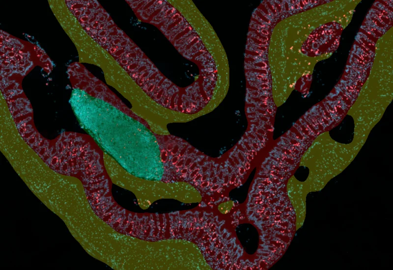
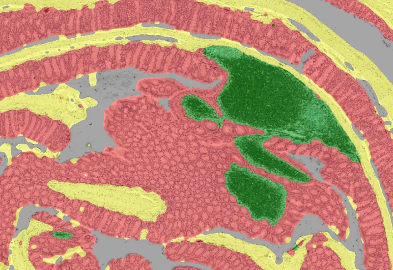
The Mucin Swiss Roll App allows for detection of the swiss roll, and the segmentation into different subclasses (mucosa, immune cell follicles, connective tissue, background). Further it detects nuclei and (e.g. PAS stained) mucin. The App outputs area (µm2) of detected tissues/tissue classes, count of total cells and in each detected area as well as the area of stained mucin in the entire tissue and within the subclasses.
Image courtesy of Priv.-Doz.Dr. Martin Schepelmann, Medical University of Vienna
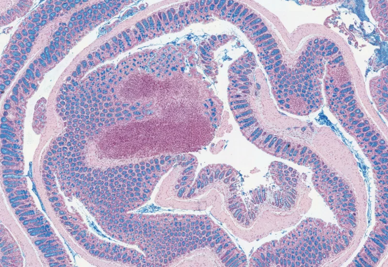
Original Image
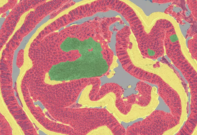
Tissue classification
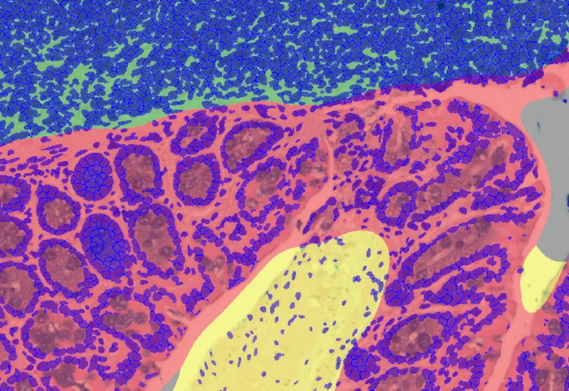
Nuclei detection
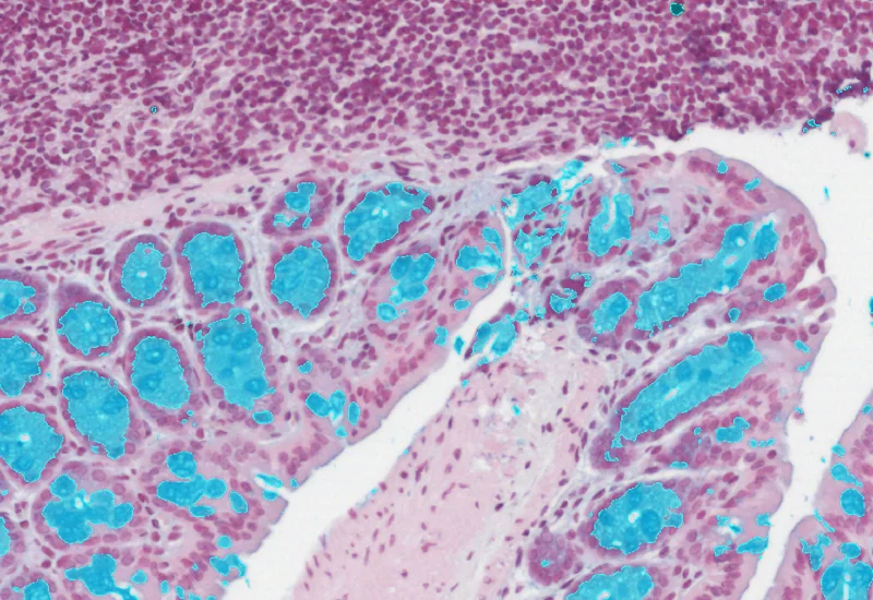
Mucin detection
single-cell analysis

Blog Post
23 Jul, 2026
How Histology Slide Scanners are Used to Study Osteoarthritis
metastructures

Blog Post
17 May, 2023
An Intro to Deep Learning in Biomedical Imaging
We support the following file formats:
- TissueFAXS (aqproj)
- StrataFAXS II (vmic)
- PreciPoint (vmic, gtif)
- Generic BigTIFF Import
- Support for multipage BigTIFF files
- OME-TIFF
- JPEG, PNG, BMP, TIFF
- Zeiss (czi)
- Hamamatsu NanoZoomer (ndpi)
- Aperio (svs)
- Leica (scn)
- 3D HISTECH Pannoramic
- Mirax (mrxs)
- Olympus (vsi)
- More slide scanners to be added!
Related Apps
IF Swiss Roll
The IF Swiss Roll App segments tissue into subclasses (e.g., mucosa, follicles, connective tissue), detects nuclei, and identifies phenotypes via IF stains.
mouse, colon, fluorescence, immune cell follicles

Custom App development
Perfectly tailored image analysis solutions for your research.
You have a specific research question that needs to be answered? We offer custom development of image analysis pipelines for specific tasks, be it detection of cellular phenotypes or quantification of tissue structures. After discussing your goals with one of our experts, you will get a ready-to-use App and be a step closer to an impactful publication.