IHC Angio Trichome
Detect trichome-stained vessels, quantify vessel, lumen, and endothelium areas, measure wall thickness and density, and analyze spatial proximity of IHC-stained cells to vessels.
trichome staining, blood vessels, collagen, muscle fibres, spatial interactions
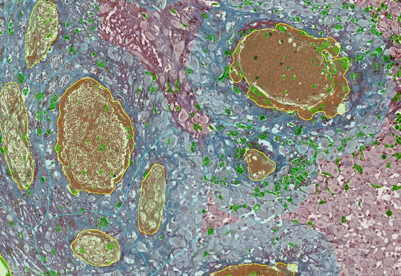
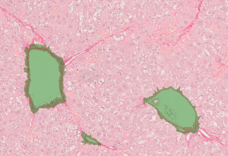
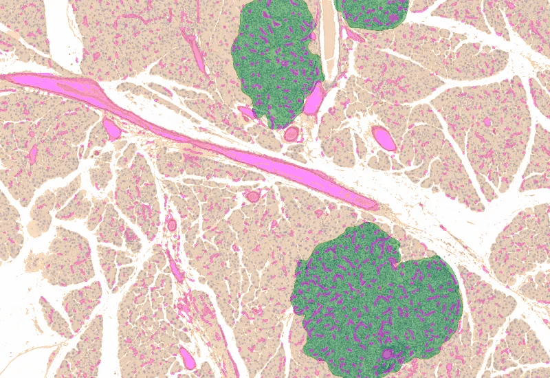
The IHC Angio Trichome App detects blood vessels based on trichome staining and measures overall vessel area as well as lumen area. Furhtermore it detects specifically IHC stained single cell populations and establishes their spatial relationship between to the detected blood vessels. The APP outputs number and vessel density, vessel wall thickness as well as areas of vessels, endothelium and lumina, number of IHC stained cells, proximity measurement etc.
Sandra Ashton, City St George's, University of London, UK

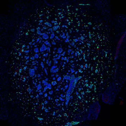
Original Image

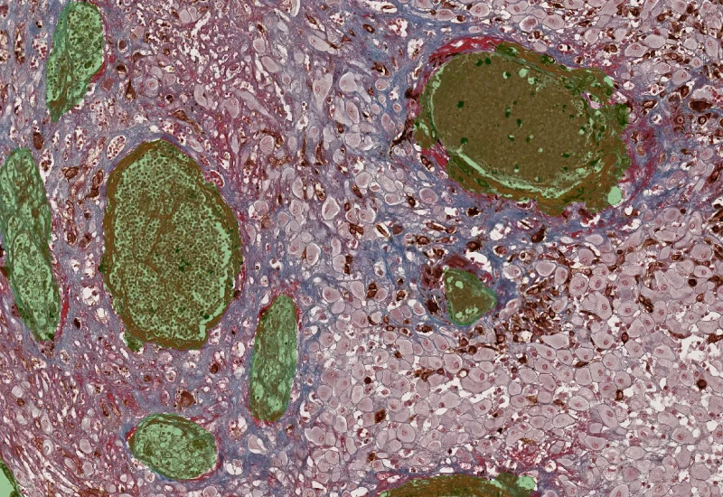
Vessel detection
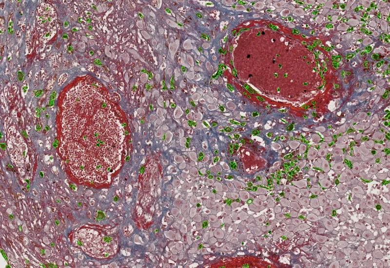
Marker positive cell detection
Combined detection
Application Note
01 Jun, 2023
Evaluating the Distance of Tumor Cells from Blood Vessels

Blog Post
17 May, 2023
An Intro to Deep Learning in Biomedical Imaging
We support the following file formats:
- TissueFAXS (aqproj)
- StrataFAXS II (vmic)
- PreciPoint (vmic, gtif)
- Generic BigTIFF Import
- Support for multipage BigTIFF files
- OME-TIFF
- JPEG, PNG, BMP, TIFF
- Zeiss (czi)
- Hamamatsu NanoZoomer (ndpi)
- Aperio (svs)
- Leica (scn)
- 3D HISTECH Pannoramic
- Mirax (mrxs)
- Olympus (vsi)
- More slide scanners to be added!
Related Apps

IHC Angio Elastin
Detect blood vessels in Verhoeff–Van Gieson–stained sections and quantify vessel number and area, as well as elastin and collagen area and intensity within user-defined perivascular rings.
blood vessels, elastine, collagen, muscle fibres, spatial interactions, Verhoeff van Geison staining
Sirius Red Angio
Detect Sirius Red–stained collagen and blood vessels, and quantify collagen area and total vessel count within the analyzed tissue.
sirius red, collagen, blood vessel, vascularization, liver

Custom App development
Perfectly tailored image analysis solutions for your research.
You have a specific research question that needs to be answered? We offer custom development of image analysis pipelines for specific tasks, be it detection of cellular phenotypes or quantification of tissue structures. After discussing your goals with one of our experts, you will get a ready-to-use App and be a step closer to an impactful publication.
