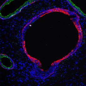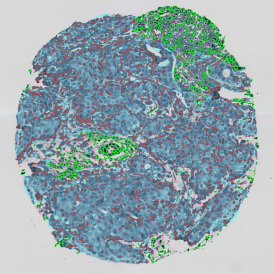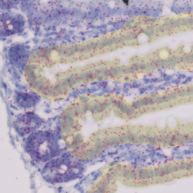


IHC Immune Status in situ APP: CD3+ cells in epithelial and interstitial tissues
The aim of the project was to quantify the distribution of CD3+ cells in and around the epithelial areas of prostate biopsies from a clinical study. The biopsies were provided by the Medical University of Vienna. The following images show some of the analysis steps.

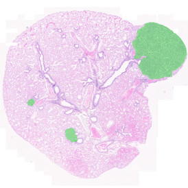
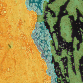
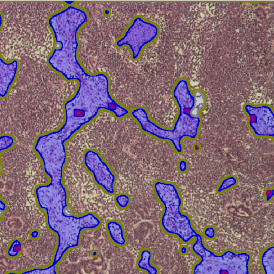
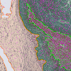
The first step in the APP is Tissue Detection.

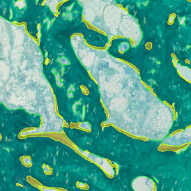
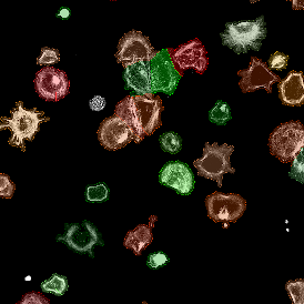
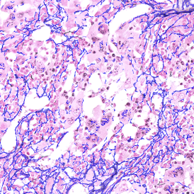
In a second step a composite image of both brown and blue channels is created.

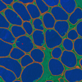
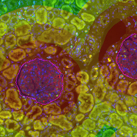
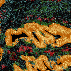
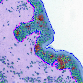
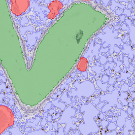
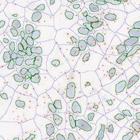
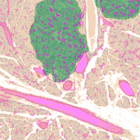
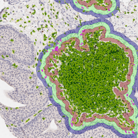
Then, a density map of the nuclei is built to determine areas with denser and larger nuclei (epithelial area candidates).

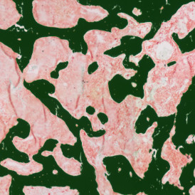
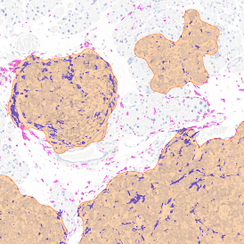
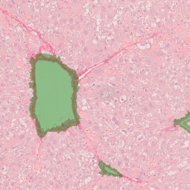
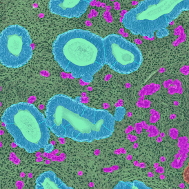
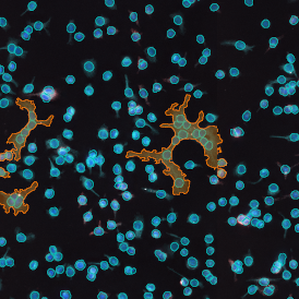
Based on the density map the epithelial areas are detected and cleaned.

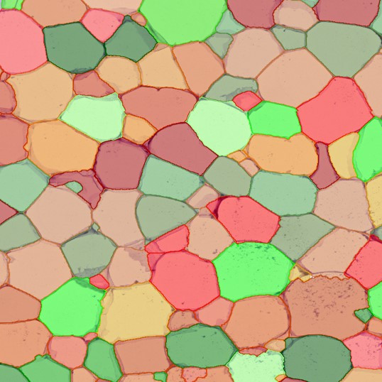
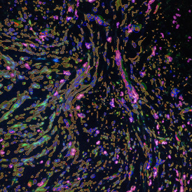
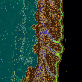
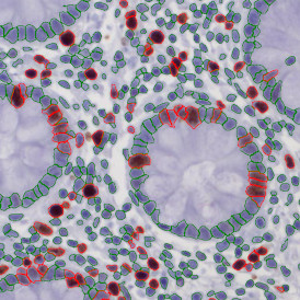
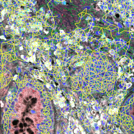
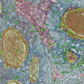
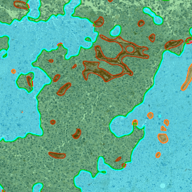
Finally, distance ranges are projected from the perimeters of the epithelial areas to provide distance measurements for CD3+ cells.

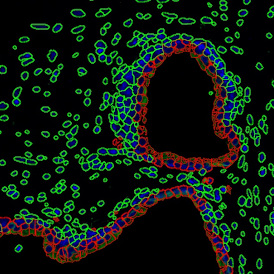
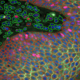
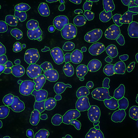
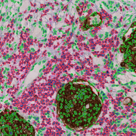
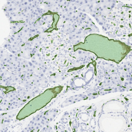
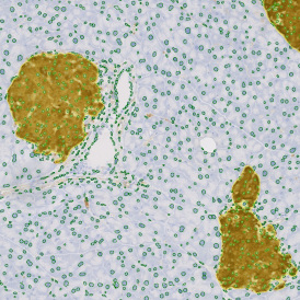
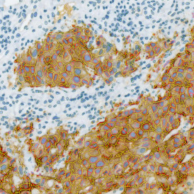
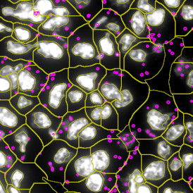
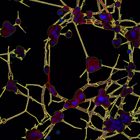
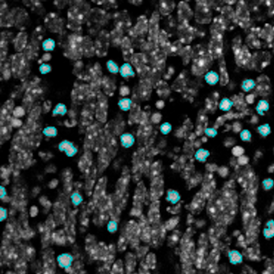
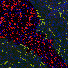
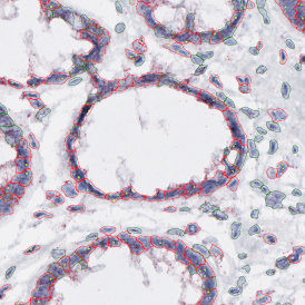
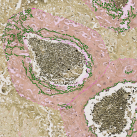
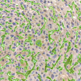
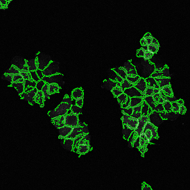
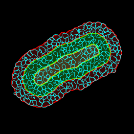
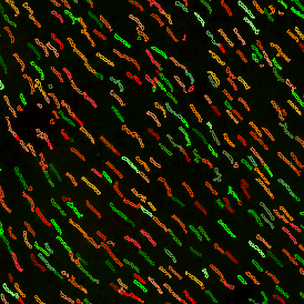
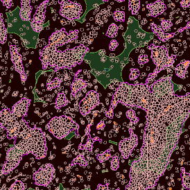
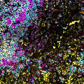
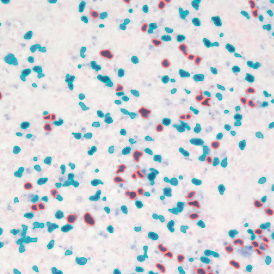
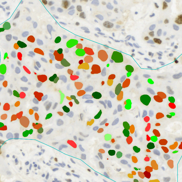
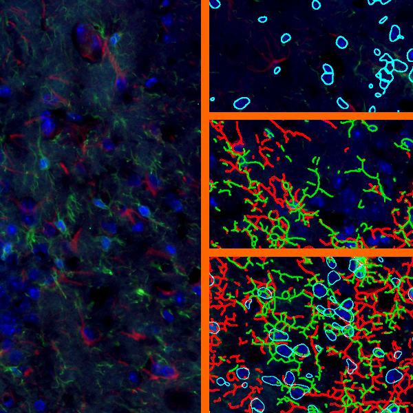
Detailed View
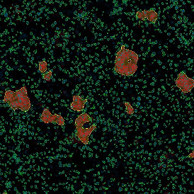
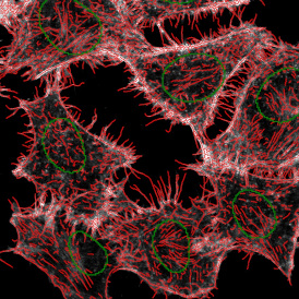
The three detailed view images below show the detection of nuclei and CD3+ staining.