Full 1
TISSUE & CELL CULTURE CYTOMETERS
TissueFAXS i PLUS
BRIGHTFIELD & FLUORESCENCE IMAGING
Full 1

TissueFAXS i PLUS 
INVERTED BRIGHTFIELD AND FLUORESCENCE SYSTEM

TissueFAXS i PLUS is an inverted fluorescence and brightfield system for the scanning and analysis of slides, cytospins, smears and Tissue Microarrays. The system is equipped with TissueQuest/HistoQuest or StrataQuest PLUS image analysis software.

S
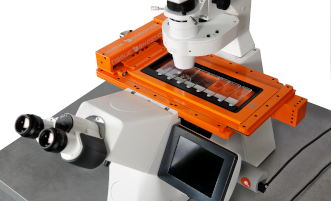
The TissueFAXS i PLUS is also available in a scan only configuration.


checkThe TissueFAXS i PLUS can be upgraded to the TissueFAXS iQ+ configuration and additionally equipped with fluorescence high-speed packages.

TissueFAXS i PLUS
KEY FEATURES

  • Multi-channel fluorescence & brightfield
  • 8 slides, 1 micro well plate, 1 petri dish
  • Automatic tissue detection
  • Time lapse scanning, live imaging
  • Extended focus, stitching & illumination correction
  • Quantitative image analysis
  • TMA & FISH/CISH scanning & analysis
  • Image and metadata storage, management and archiving
  • Default
  • Title
  • Date
  • Random

CONFIGURATION

TissueFAXS i PLUS is the most versatile configuration of all TG inverse systems. It can scan and analyse samples on slides, in microtiter plates and in Petri dishes in both brightfield and epifluorescence modes. It can be equipped with supplementary components for contrast microscopy methods, like Differential interference contrast (DIC) microscopy in brightfield. It scans 5 channels in epifluorescence if the maximum of 5 single bandpass filters is fitted or more if multibandpass filters are used.

The system comes equipped with an 8-slide stage which can also take a microtiter plate insert as an alternative. Inserts for Petri dishes and customized inserts for other sample containers are available on demand. A four slide/one microtiter plate stage is also available.

TissueFAXS i PLUS supports Live Imaging, as do all other inverse stand-based configurations. CO2, O2 and temperature control modules are available, as is the choice of small stage top incubators or conventional incubation enclosures. The environment controls are not integrated into the TissueFAXS control software (being usually set for longer time periods) and can be controlled via the microscopes touch screen. The TissueFAXS control software provides time lapse scanning.

Objectives from all Zeiss objective classes with M24 thread can be fitted, the turret takes a maximum of 6. TG continuously checks the market for cameras and other technical equipment to provide those solutions that deliver top performance and are cost-effective. The TissueFAXS i PLUS standard cameras are good examples of this successful strategy. The right illumination is of paramount importance in light microscopy. TG provides fully system software-controlled solutions which are mostly maintenance free and have long lasting illuminantes. The correct filters which guarantee pixel shift-free and low channel crosstalk are equally important to make fluorescence scanning easily accessible for all users of a TG system.

TissueFAXS Scanning and Management software integrates all the hardware components into an easy to use workflow with full, walk-away automation capabilities.


TissueFAXS inverted Configuration

TissueFAXS iPlus LiveImaging

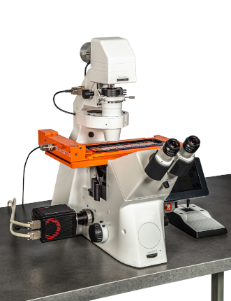
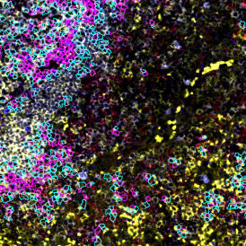
Adipocyte App: Measurement of Cellular Size of Adipocytes

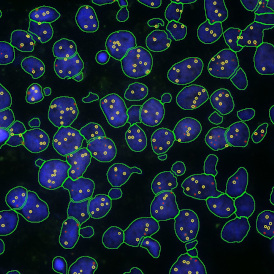
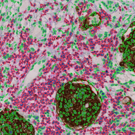
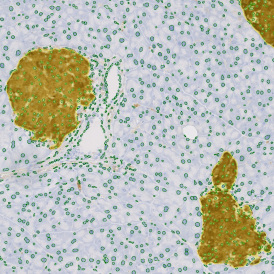
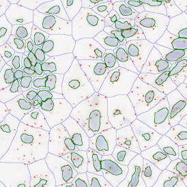
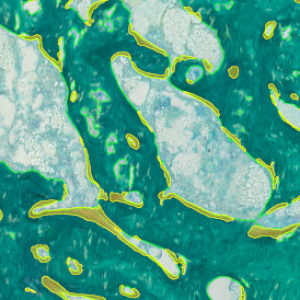
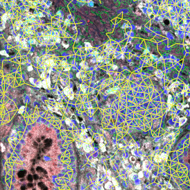
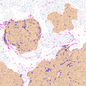
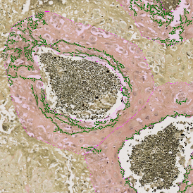
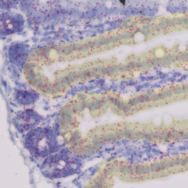
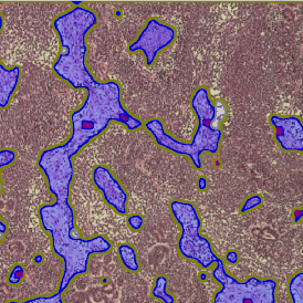
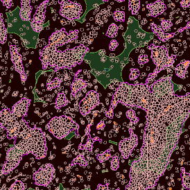
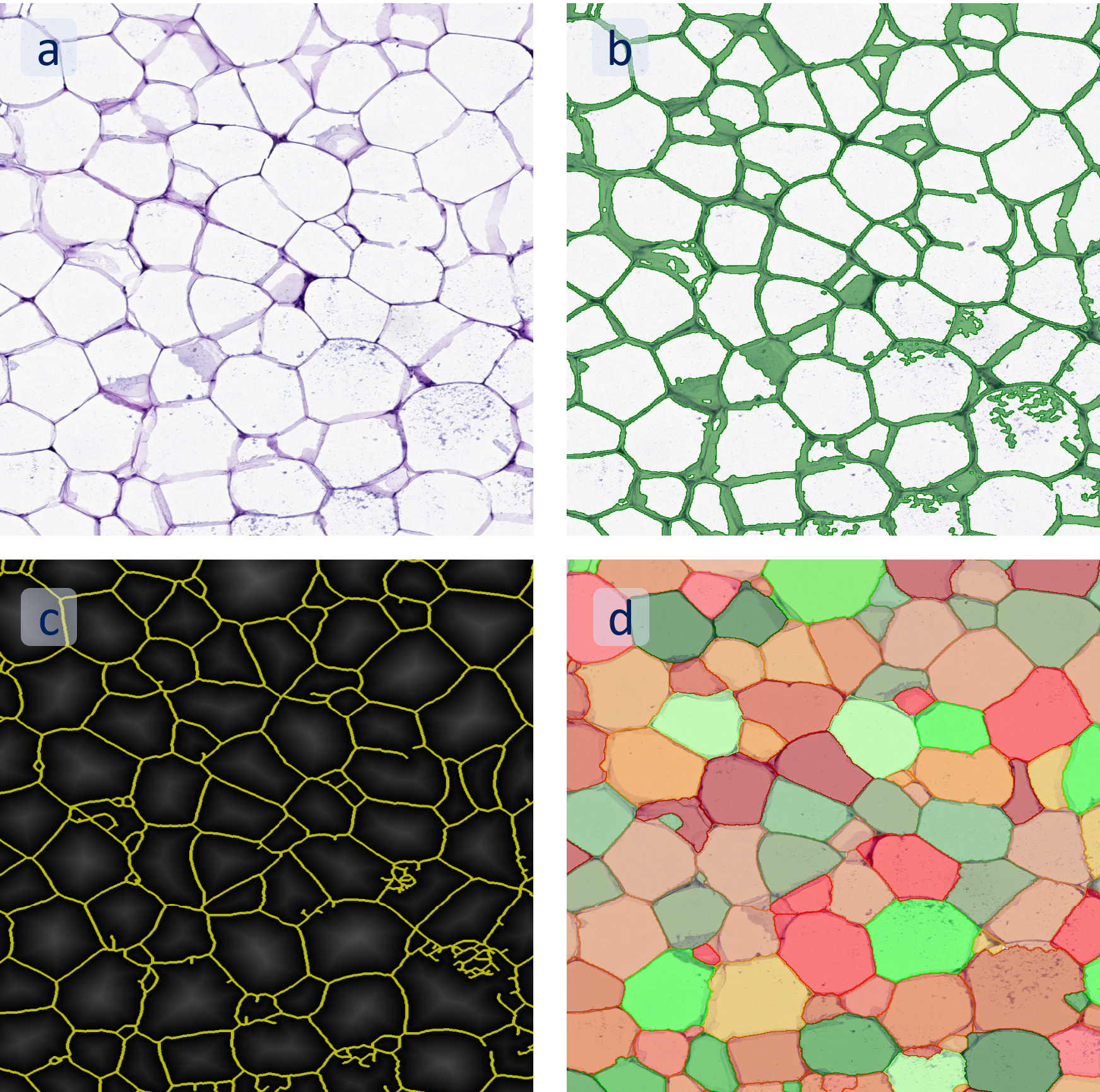
The StrataQuest-based Adipocyte App was applied to this sample with the aim to automatically detect (i) the number, (ii) the size, and (iii) the diameter of adipocytes within the given fat tissue. The challenge of analyzing the adipocytes is that the nuclei cannot be detected properly since they are either usually washed away during the H&E staining process, or are not visible. Therefore, the App detects the adipocytes using the membranes of the cells.

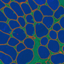
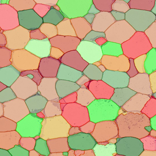
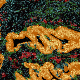
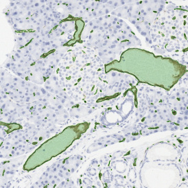
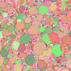
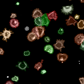
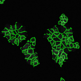
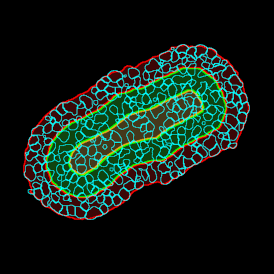
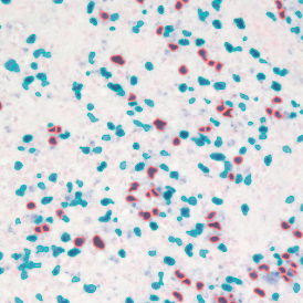
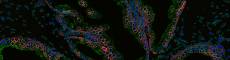
(a) Shows fat tissue stained with hematoxylin & eosin (H&E). The App first detects the membranes of the adipocytes and minor membrane tears are automatically closed (b).  To detect the individual cells without nuclei, the App uses the detected membranes as a starting point and defines the cell by its distance from the membrane (c). Here the final result of cell detection is shown (d), cells are labeled in different colors for visualization purposes.

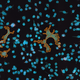
The App automatically eliminates adipocytes on the borders from the analysis which may influence the proper outcome of the analysis.

 IF_Tumor_Foci_Angio_overview-min.png

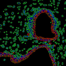
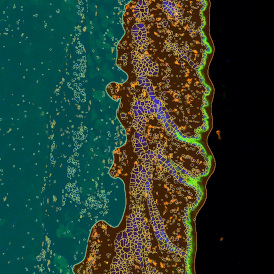
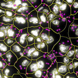
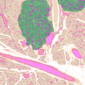
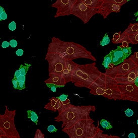
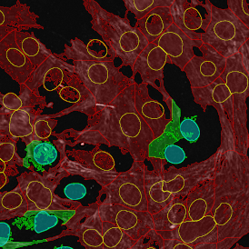
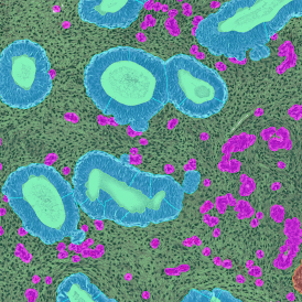
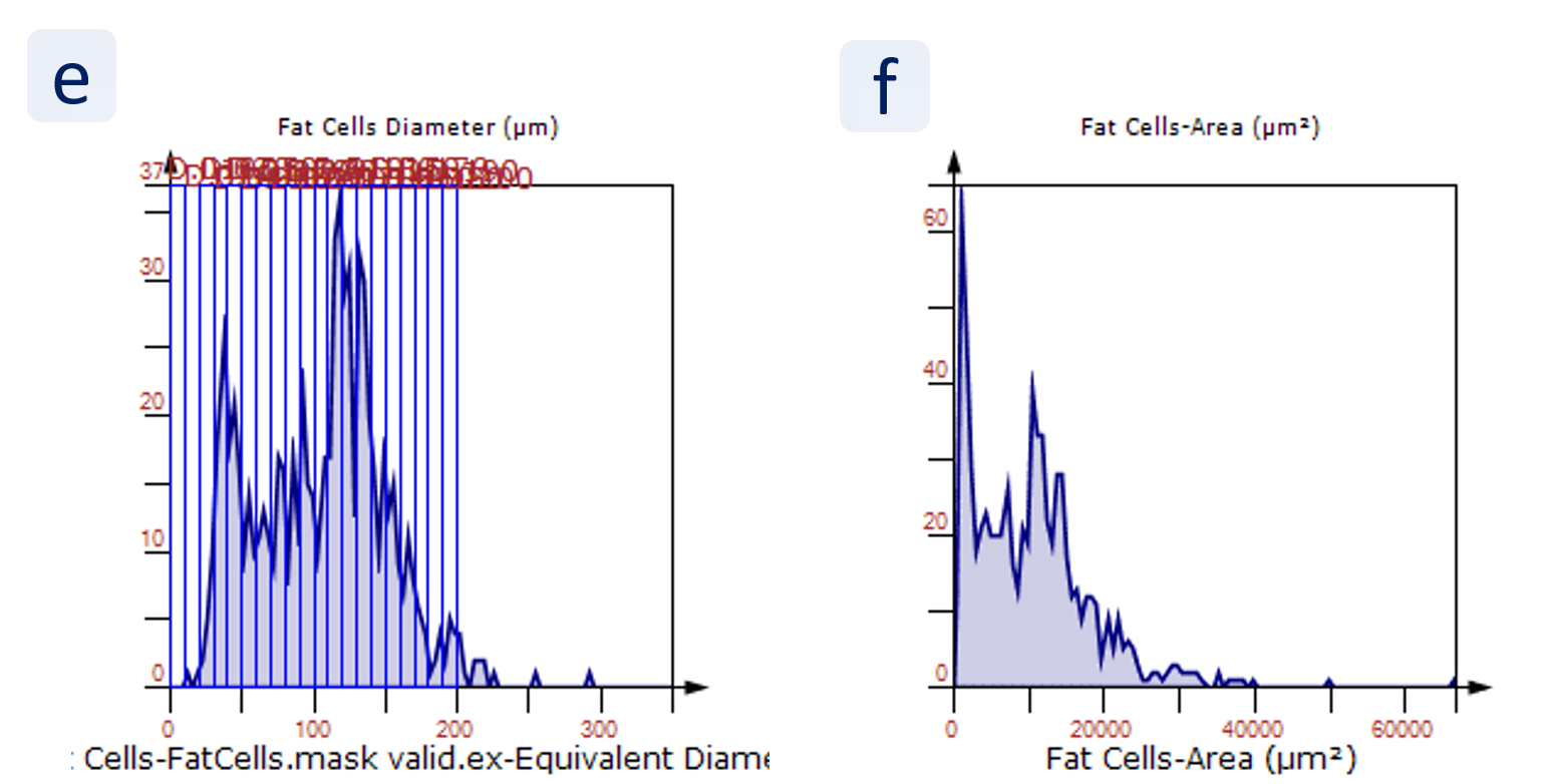
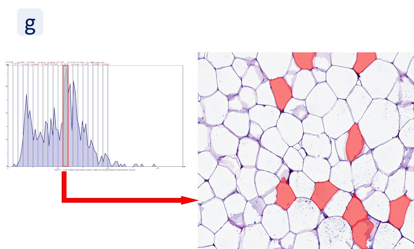
The results of the analysis are visualized in histograms and scattergrams. As shown in the scattergram below, the cells can be grouped according to their diameter (e) or (f) size.

IF_Tumor_Foci_Angio_panel-min.png

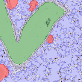
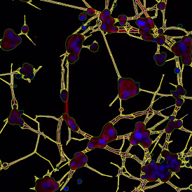
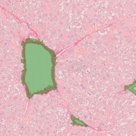
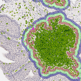
By selecting a gate of interest within the scattergram and using the backward connection feature, the cells from the selected gate are viewed directly in the sample in their native tissue environment (g). In this case, cells with diameters of 120µm to 129µm are shown in red.

IF_Tumor_Foci_Angio_scattergram.png

The gained measurements can be exported from StrataQuest in CSV and /or Excel format.

StrataQuest Apps offer a streamlined and easy workflow for analyzing your samples. Look through our StrataQuest Apps and get inspired by the variety of biomedical image analysis applications. If you cannot find the perfect solution for your research question, please contact us. TissueGnostics offers the development of customized Apps for your specific analysis needs.

PROPERTIES

Design

Inverted microscope, modular (Microscope, Cameras, Light-source, PC with 2 Monitors)

Microscopy mode

Fluorescence and brightfield scanning

Compatible Slide Formats

Standard Slides, Over-sized Slides, Petri Dishes, Well-Plates

Slide Capacity

8

Objectives

Up to 6 Objectives (2.5-100x)

FL-Channels

5

Camera

Monochrome: sCMOS camera (16-bit, 2048x2048px)
Color: CMOS camera (8-bit, 2048x2048px)

Light Source

VIS-LED
Solid-state Light Engine

Tissue Microarray (TMA)

Yes

Image Analysis Software

contextual image analysis - StrataQuest, StrataQuest Apps
single cell analysis - TissueQuest and HistoQuest

Supported Image Formats

TissueFAXS, OME-TIFF, TIFF, JPEG, BMP, PNG

Viewer

TissueFAXS Viewer: integrated, stand-alone freeware

Contact

TissueGnostics GmbH
Taborstraße 10/2/8
1020 Vienna, Austria
+43 1 216 11 90
This email address is being protected from spambots. You need JavaScript enabled to view it.

We use cookies on our website. Some of them are essential for the operation of the site, while others help us to improve this site and the user experience (tracking cookies).
You can decide for yourself whether you want to allow cookies or not. Please note that if you reject them, you may not be able to use all the functionalities of the site.