• 1

WELCOME

TissueGnostics provides fully integrated cutting-edge tissue cytometers for (i) whole-slide imaging (brightfield, fluorescence, confocal, multispectral) and (ii) high-end analysis of tissue sections, cultured cells, TMAs, smears etc. Explore TGs solutions for spatial phenotyping, single cell analysis, molecular single cell profiling, machine-learning based tissue classification and many more.
 
IF COEXPRESSION APP

IHC META.CELLS

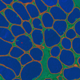
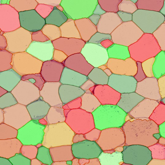
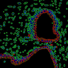
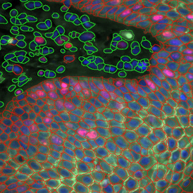
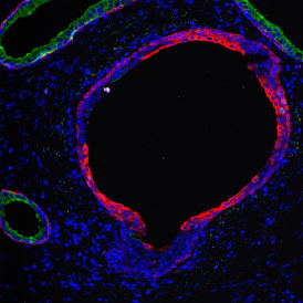
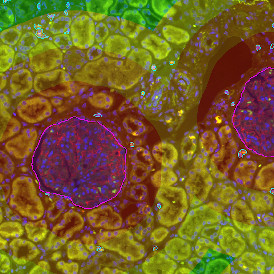
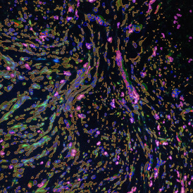
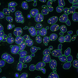
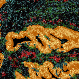
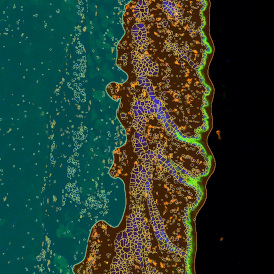
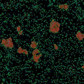
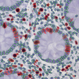
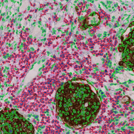
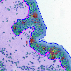
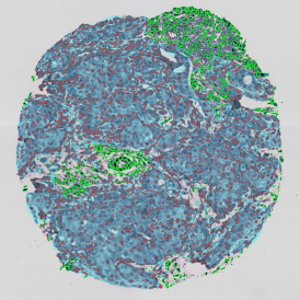
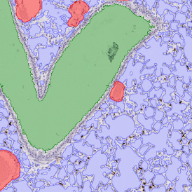
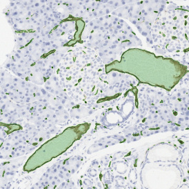
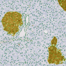
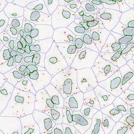
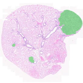
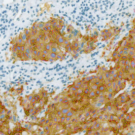
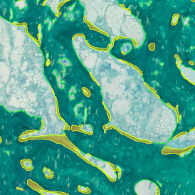
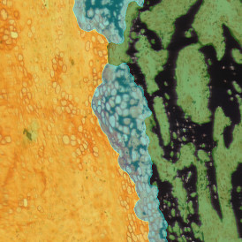
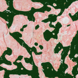
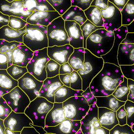
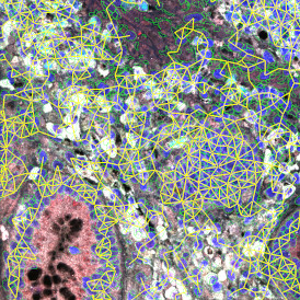
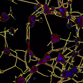
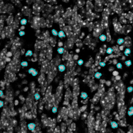
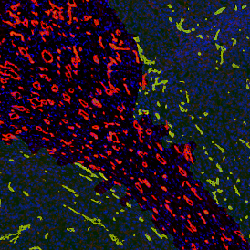
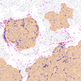
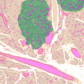
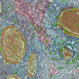
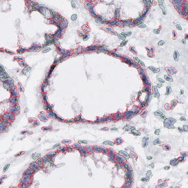
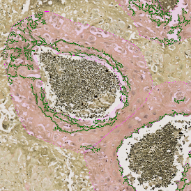
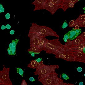
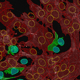
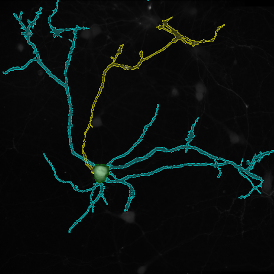
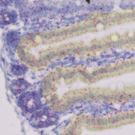
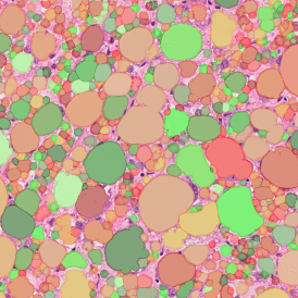
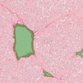
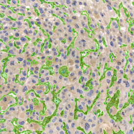
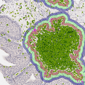
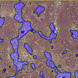
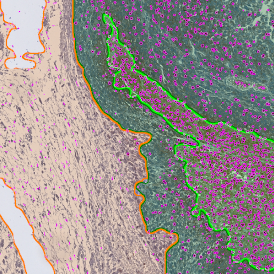
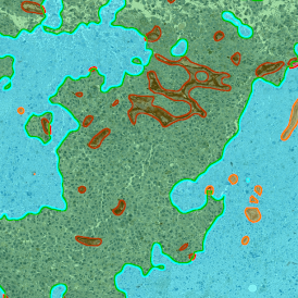
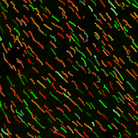
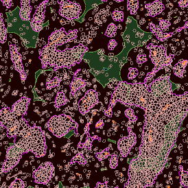
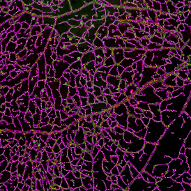
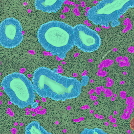
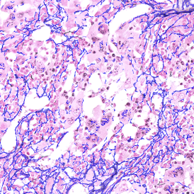
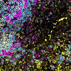
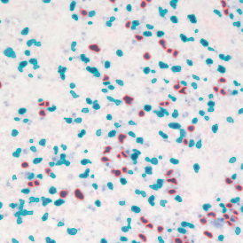
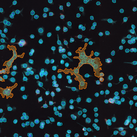
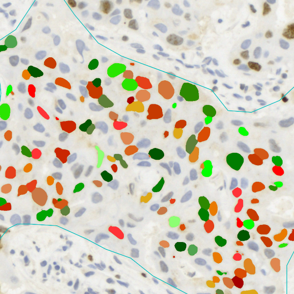
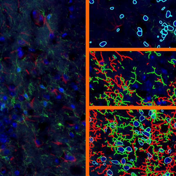
The IHC Meta.Cells APP combines the detection of IHC/HC stained metastructures (e.g. Langerhans islets) with single cell detection (segmentation of cells into nucleus, and/or perinuclear area and/or cytoplasm). Detected cells can be classified and visualized as being either within or outside of detected metastructures. Each detected area and cell compartment is measured for up to 20 intensity, statistic and morphometric parameters.

APP Category 2


IF COEXPRESSION APP

IHC IMMUNE STATUS IN SITU

The IHC Immune Status in Situ APP provides phenotypic characterization of immune cells in context with detected metastructures (e.g. tumors, glands). It measures the distance of cellular objects to the metastructure boundary (within and/or outside). Distance ranges can be defined. Each segmented cell compartment is measured for up to 20 parameters, as is the distance of each cell to the boundary.

APP Category 3

IF COEXPRESSION APP

IHC3

The IHC3 APP unmixes three markers (e.g. two chromogens and counterstain) in an IHC or HC digital slide and segments single cells into nucleus, and/or perinuclear area and/or cytoplasm. Each segmented cell compartment is measured for up to 20 intensity, statistic and morphometric parameters which are displayed in scattergrams and histograms and can be exported.

APP Category 2

IF COEXPRESSION APP

IHC TUMOR-STROMA

The IHC Tumor-Stroma APP combines the segmentation of tumor and stroma (based on the morphology) and the detection of specifically stained cell populations. It segments the cells into nucleus, and/or perinuclear area and/or cytoplasm. Each segmented cell compartment in tumor and/or stroma is measured for up to 20 intensity, statistic and morphometric parameters which can be displayed in scattergrams and histograms and exported.

APP Category 2

IF COEXPRESSION APP

IHC MULTI-SHADES

The IHC Multi-Shades APP provides semi-automatic color separation for up to six markers or colors in an IHC or HC digital slide. In the above sample it has been used to detect and segment different levels of ossification based on Azan stain. Each detected area is measured for up to 20 intensity, statistic and morphometric parameters.

APP Category 1


IF COEXPRESSION APP

RNA Scope

The RNA Scope APP provides dot detection per cell within the nuclear compartment (nucleus and/or cytoplasm) for up to three markers in CISH and SISH experiments. Each segmented cell compartment is measured for up to 20 intensity, statistic and morphometric parameters. Dot parameters are provided per cell and include count, mean intensity, total dot area, and sum of intensity as well as area and intensity lists for all single dots.

APP Category 2

IF COEXPRESSION APP

CLASSIFIER

The Classifier APP is based on trainable Deep Learning technology and provides tissue metastructure detection. In the above example it separates the colon tissue into crypts, stroma and infiltration areas. Combined with the IHC2 cellular detection APP the Ki-67+ nuclei in each compartment can be quantitatively analyzed for up to 20 parameters.

APP Category 3

IF COEXPRESSION APP

TUMOR FOCI

The Tumor Foci APP allows to detect the whole tissue and more important tumor foci based on nuclear structure analysis, mainly on HE staining. The number and area of tumor foci as well as their density is measured. 

APP Category 1

IF COEXPRESSION APP

IHC²

The IHC² APP unmixes two markers (e.g. chromogen and counterstain) in an IHC or HC digital slide and segments single cells into nucleus, and/or perinuclear area and/or cytoplasm. Each segmented cell compartment is measured for up to 20 intensity, statistic and morphometric parameters which are displayed in scattergrams and histograms and can be exported. 

APP Category 1


IF COEXPRESSION APP

IHC Angio

The IHC Angio APP detects blood vessels based on appropriate stains (e.g. CD31) and measures overall vessel area as well as lumen area. The vessel detection also can be set to close open stained vessel walls and to connect separated vessel sections within a definable distance. The APP outputs number and vessel density as well as areas of vessels, endothelium and lumina.

APP Category 2

IF COEXPRESSION APP

IHC ADIPOCYTE

The IHC Adipocyte APP quantifies adipocytes as to their lumen in adequate HE samples. Small rips in adipocyte membranes are mended automatically and cell membrane artefacts in adipocyte lumina are automatically eliminated as are lumina on sample borders. The APP outputs area measurements for all detected adipocyte lumina.

APP Category 1

IF COEXPRESSION APP

PULMO

The Pulmo APP segments the metastructure components of lung, including tissue, bronchioles, blood vessels and alveoles. Each segmented metastructure is measured for up to 20 morphometric parameters.

APP Category 3

IF COEXPRESSION APP

IHC Membrane

The IHC Membrane APP unmixes up to three markers in an IHC or HC digital slide and segments cells into nucleus, and/or perinuclear area and/or cytoplasm, as well as into membrane (e.g. HER2/neu). Each segmented cell compartment is measured for up to 20 intensity, statistic and morphometric parameters. Three more parameters are measured for membrane intensity and angle of staining. All parameters are displayed in scattergrams and histograms and can be exported.

APP Category 2

IF COEXPRESSION APP

IHC MACROPHAGES

The IHC Macrophages APP detects macrophages based on adequately stained IHC samples. The APP can be combined with area detection and distance range algorithms, in the sample above to determine the distance of Langerhans cells from the border of the epidermis within and without. Each segmented cell compartment is measured for up to 20 parameters, as is the distance of each cell to the boundary.

APP Category 3

 
  • TG AUSTRIA
    Global Headquarter

    Taborstrasse 10/2/8
    A-1020 Vienna
    AUSTRIA, EU

    Tel.: +43/1/216 11 90

    This email address is being protected from spambots. You need JavaScript enabled to view it.


    TG ROMANIA

    Str. Sf. Andrei, nr.15A
    700028 Iasi
    ROMANIA, EU

    Tel.: +40/332/40 58 66

    This email address is being protected from spambots. You need JavaScript enabled to view it.

    Read More
  • TG USA 

    12522 Moorpark Street
    Suite #106
    Los Angeles, CA 91604
    USA

    Tel.: +1/818/856/8056

    This email address is being protected from spambots. You need JavaScript enabled to view it.


    TG USA East

    ScientiaLux d.b.a.
    4 Farnum Terrace
    Worcester, MA 01602
    USA

    Tel.: +1/508/471/7732

    This email address is being protected from spambots. You need JavaScript enabled to view it.

    Read More
  • TG ASIA PACIFIC

    China Division
    Room 506,
    No.6 Auto Museum East Rd,
    Fengtai District Beijing
    CHINA

    Tel.: +86/400/898 1980

    This email address is being protected from spambots. You need JavaScript enabled to view it.


    Western Pacific Division
    Taipei

    Tel.: +886/928/899 397

    This email address is being protected from spambots. You need JavaScript enabled to view it.


    Australia Division
    Brisbane, Queensland

    Tel.: +61/416 037 618

    This email address is being protected from spambots. You need JavaScript enabled to view it.

    Read More
  • TG AFRICA

    Measuring Instruments Technology (MIT)
    CSIR Campus, Building 33
    Meiring Naude Rd
    Brummeria, Pretoria 0181
    SOUTH AFRICA

    Tel.: +27 12 349 5191
    Cell: +27 82 784 2011

    This email address is being protected from spambots. You need JavaScript enabled to view it.

    Read More
  • 1

Contact

TissueGnostics GmbH
Taborstraße 10/2/8
1020 Vienna, Austria
+43 1 216 11 90
This email address is being protected from spambots. You need JavaScript enabled to view it.

We use cookies on our website. Some of them are essential for the operation of the site, while others help us to improve this site and the user experience (tracking cookies).
You can decide for yourself whether you want to allow cookies or not. Please note that if you reject them, you may not be able to use all the functionalities of the site.