APP SHOWCASE
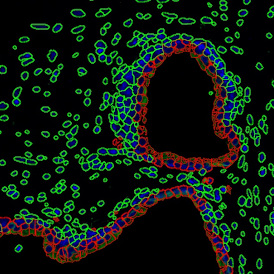
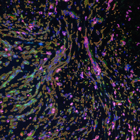
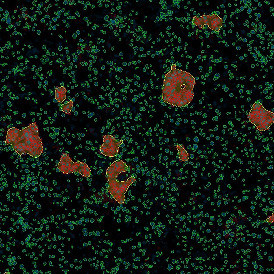
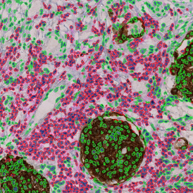
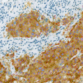
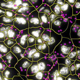
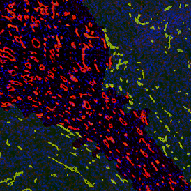
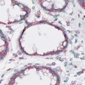
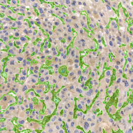
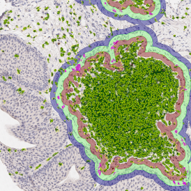
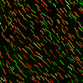
The IF Cellular Microenvironment App allows to determine the cellular phenotype of specific IF-stained cell populations.

The IF Cellular Microenvironment App allows to determine the cellular phenotype of specific IF-stained cell populations.
TG ACADEMY WEBINAR

Dr. Henning Ulrich
University of Sao Paulo

2023 Center of Excellence awards are given to celebrate and acknowledge our most impactful academic partnerships.

In research, techniques like immunofluorescence staining combined with tissue cytometry are crucial for studying biology.

A recent study revealed important information on how cytolytic CD8+ T cells are central in determining long-term viral control.
Explore our online database of reference publications


Dr. Rupert Ecker, CEO of TissueGnostics, has achieved a remarkable milestone by gracing the cover of the World's Leader Magazine.

REDI Fellow Jyotsna Batra successfully completes her fellowship with TissueGnostics to battle prostate cancer.


Explore our analysis solutions in our Strataquest Appcenter and get inspired by the variety of applications.
adfdfasdf
The IF Cellular Microenvironment App allows to determine the cellular phenotype of specific IF-stained cell populations.
TG ACADEMY WEBINAR
Dr. Henning Ulrich
University of Sao Paulo
2023 Center of Excellence awards are given to celebrate and acknowledge our most impactful academic partnerships.
In research, techniques like immunofluorescence staining combined with tissue cytometry are crucial for studying biology.
A recent study revealed important information on how cytolytic CD8+ T cells are central in determining long-term viral control.
Explore our online database of reference publications
Dr. Rupert Ecker, CEO of TissueGnostics, has achieved a remarkable milestone by gracing the cover of the World's Leader Magazine.
REDI Fellow Jyotsna Batra successfully completes her fellowship with TissueGnostics to battle prostate cancer.
Explore our analysis solutions in our Strataquest Appcenter and get inspired by the variety of applications.
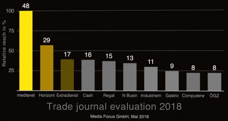
22. Nov 2018
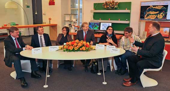
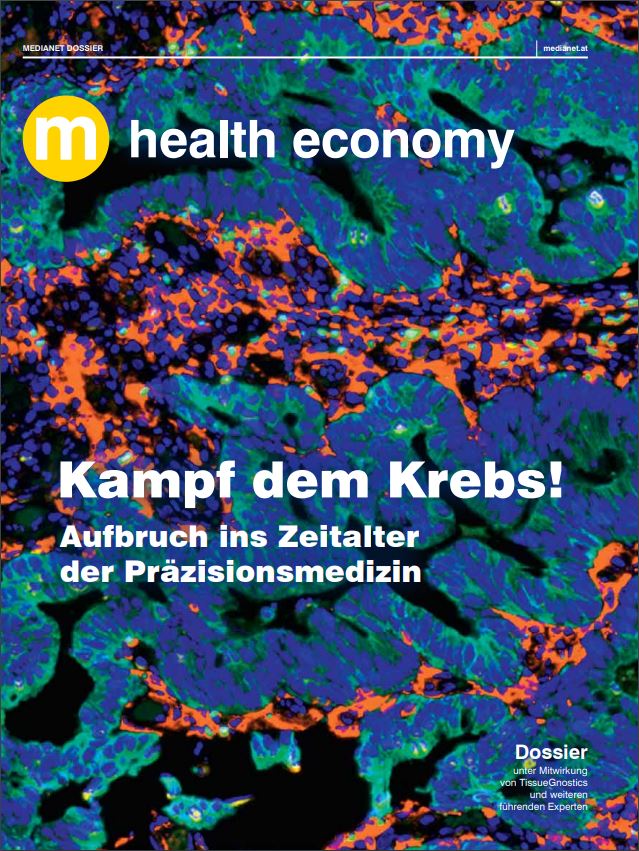
On October 5th 2018 the Austrian Medianet magazine published a health economy dossier on Personalized Medicine.
Medianet has the largest reach of all Austrian trade journals and is mainly read by managers, decision makers and stakeholders in the Austrian economy.
TissueGnostics globally contributes to research which boosts Personalized Medicine through cooperation projects and enabling research done by its clients. Therefore the company was invited to organize a round table on the subject with Austrian experts and also to provide further insights from its cooperation partners.
The round table was held at TissueGnostics premises with TGs CEO, Rupert Ecker, taking part as an expert.
The dossier will be included in the medianet print journal, with a circulation of 14.000 and as an e-paper with a circulation of 42.000 .
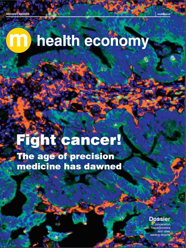
Download the pdf version of the dossier.
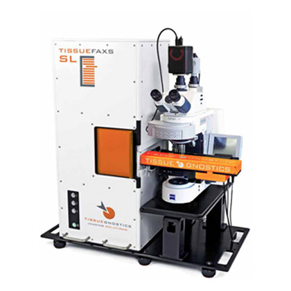
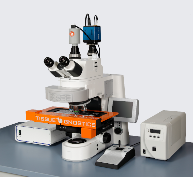
TissueGnostics (TG) provides streamlined solutions for both biomedical imaging and image analysis. The goal of TG is to bring the same type of phenotypical analysis of single cells from Flow Cytometry into tissue context. The merging of image analysis software and high-quality optics and robotics has allowed TG to create the TissueFAXS system - a fully automated system which can scan slides/well plates and automatically quantify marker expression per cell. Going a step beyond that is StrataQuest - a software development platform for creating complex image analysis algorithms that can automatically detect multicellular structures within scanned tissue sections for a highly detailed contextual tissue analysis.
The imaging systems are modular and upgradable. Every system can be customized to offer the following capabilities: brightfield scanning, widefield fluorescence, confocal, and multispectral. Every system can come either in an upright configuration for scanning slides only, or inverted for scanning well plates and slides. Each TissueFAXS system comes with either an 8-slide stage, or a high-throughput automatic 120 slide loading system only available for the upright TissueFAXS system configuration. TissueFAXS systems can come with high powered LED light engines combined with multi bandpass filter cubes for high speed fluorescence scanning.
Image analysis software comes in 3 forms: TissueQuest for fluorescence image analysis, HistoQuest for brightfield image analysis, and StrataQuest for multicellular contextual tissue analysis for both brightfield and fluorescence images. TissueQuest and HistoQuest are streamlined for rapidly acquiring nuclear segmentation and marker quantification per cell. StrataQuest offers much more in terms of image analysis and is therefore more complex, which is why TissueGnostics offers the development of customized algorithms as a service to researchers. Every StrataQuest solution (or APP) includes a simplified user interface that is made by the underlying algorithm, and contains macros so that even researchers with little or no experience in image analysis can obtain high quality data from analysing their scanned images.
TG provides image analysis solutions for a multitude of research questions. The image analysis software StrataQuest, HistoQuest and TissueQuest (for brightfield and fluorenscence projects) can be applied e.g. to explore the tumor microenvironment and/or the spatial organization of cellular subpopulations, to detect and quantify fluorescence in situ hybridization (FISH), to assess different bone structures, or to analyze multiplex IF stainings. Please find additional applications in the sections: App center and user examples in StrataQuest APP analysis examples.
In addition to identifying and characterizing the morphology of individual cells, a more complex phenotypic profile includes the cell’s activation state and function within the tissue microenvironment.
Recent developments in quantitative imaging hardware and software have allowed researchers to study immune cells in situ but, as only four biomarkers can be studied at one time on standard microscopy systems, this does not capture a complete picture of the cell’s activity.
Meanwhile, flow cytometry is a well-established method that allows multiple biomarkers to be studied at one time, but it has the disadvantage of dissociating the cells from important information about the tissue microenvironment.
The development of a system that could provide flow cytometry-like data on cell biomarkers while retaining information on the tissue microenvironment, as in conventional microscopy, would therefore be extremely valuable for meeting the demands of modern clinical practice.
A recent study by researchers at Yale University School of Medicine (Blenman K & Bosenberg M, 2019) demonstrates an approach that can overcome the limitations discussed above. They show that applying new analytic technology to a conventional microscopy setup makes possible a complex characterization of immune cells in situ.
Adapting a familiar setup
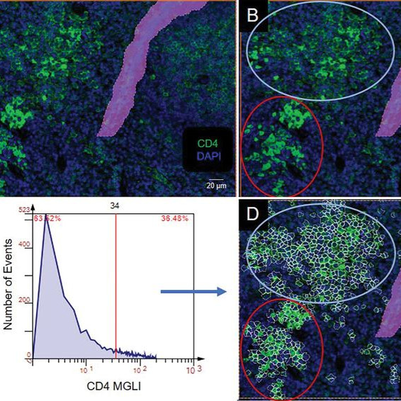
Dr Kim Blenman studied tissue samples from the spleen in a mouse model of melanoma developed by Dr Marcus Bosenberg. Dr Blenman employed a conventional microscopy setup to perform multiplexed fluorochrome-based histology on formalin-fixed paraffin-embedded tissue sections, with biomarkers conjugated to Cy3 or Cy5.
Dr Blenman was interested in the CD4 transmembrane protein – a biomarker of multiple T-cell subtypes – and six biomarkers of cell activity, such as proliferation and transcription, associated with these cells.
She used multiple staining rounds, inactivating the dyes between each round, to study the different biomarkers of cell activity including Foxp3, RORγ(t), Ki-67, T-bet, Granzyme B, and IL-6.
Dr Blenman used an all-in-one system from TissueGnostics. High-magnification images were acquired with the TissueFAXS Quantitative Imaging System for downstream analysis integrated with advanced image processing software (TissueGnostics StrataQuest) that reconstructs whole images in silico and performs cell phenotyping and tissue cytometry.
The software first uses and algorithm to stitch the tiled images together to recreate the whole image. It then creates a composite image that includes all the biomarkers from each staining round. Following this, cell isolation is performed using two algorithms: one to identify the cell nucleus and a second which identifies the biomarker for the cell phenotype. Quantification was then achieved by assessing in turn which cells were positive for each biomarker, using a backgating algorithm to determine cutoffs at which cells or cell clusters should or should not be included.
Using this method on the spleen tissue samples, Dr Blenman was able to show that it can obtain multiple biomarkers at one time in situ.
With this information, they were able to phenotypically characterize cells within the tissue sample and assign them to categories based on the combination of biomarkers expressed by individual and clusters of cells. Through this, they showed that levels of CD4 expression within cell clusters correlated with distinct patterns of biomarker expression and cell proliferation.
Visualizing the future
The immune system is incredibly complex and being restricted to profiling only four biomarkers per cell type has been a limiting factor. One advantage of the system used by the authors is that it harnesses a microscopy set up that is already present in most laboratories, making it highly accessible. And, while the authors chose to study six biomarkers, there is theoretically no limit to the number of biomarkers that can be studied at one time.
Dr Blenman says that this method has an advantage of being able to objectively quantify cell biomarkers. Reporting in Cytometry Part A, she says it has potential to be very useful in cancer treatment where therapies are becoming increasingly targeted to the individual’s disease and its underlying mechanism. For example, the technique could be used to detect response to immunotherapy through the analysis of biopsies before, during, and after treatment.
The method helps to retain spatial information about the cells while characterizing them phenotypically, facilitated by flow cytometry-like capabilities including gating, backgating and histogram/dot scatterplot outputs.
TissueGnostics innovation
The process described by Dr Blenman in the Cytometry A paper was made possible through the TissueFAXS Quantitative Imaging System and TissueGnostics StrataQuest analysis software. These technologies combined lead to a streamlined, highly automated process which frees the user from hands-on time. The authors highlight the potential of TissueFAXs to incorporate MATLAB scripts into the StrataQuest analysis software. There are also over 50 compatible ready-to-load apps which can facilitate specialized analyses. Each app provides a specific analysis from start-to-finish and deliver the data ready to export to the program of your choice.
The TissueFAXS system is a versatile upright system which can scan and analyze slides, cytospins, smears and tissue microarrays. It comes equipped with both fluorescence and brightfield modes but, with supplemental components, can also be customized for contrast microscopy methods.
The system can be equipped with one of three software choices, the most advanced and comprehensive being StrataQuest. StrataQuest can automatically detect structures within a digital slide, such as blood vessels and can identify and quantify millions of cells within a cell sample. The software is also available as a standalone products for use with existing scanning systems and is compatible with imported images and slides in a wide range of formats.
References
Watch the Kim Blenman's Webinar - Practical approaches for using tissue cytometry for clinical and research applications
Blenman KRM & Bosenberg MW. Immune Cell and Cell Cluster Phenotyping, Quantitation, and Visualization Using In Silico Multiplexed Images and Tissue Cytometry. Cytometry A. 2019; 95:399-410. doi: 10.1002/cyto.a.23668.
This Privacy Policy clarifies the nature, scope and purpose of the processing of personal data (hereinafter referred to as "data") within our online service and the related websites, features and content, as well as external online service, e.g. our social media profiles on Facebook, LinkedIn, Twitter and XING (collectively referred to as "online offer"). With regard to the terminology used, e.g. "Processing" or "Responsible", we refer to the definitions in Article 4 of the General Data Protection Regulation (GDPR).
TissueGnostics GmbH
Taborstraße 10/2/8
A-1020 Vienna
This email address is being protected from spambots. You need JavaScript enabled to view it.
http://www.tissuegnostics.com/imprint
- Inventory data (e.g., names, addresses).
- contact information (e.g., e-mail, phone numbers).
- content data (e.g., text input, photographs, videos).
- usage data (e.g., websites visited, interest in content, access times).
- Meta / communication data (e.g., device information, IP addresses).
Visitors and users of the online service (hereinafter we refer to the affected persons as "users").
- Provision of the online service, its functions and contents.
- Answering contact requests and communicating with users.
-Safety measures.
- Audience measurement / Marketing
"Personal data" means any information relating to an identified or identifiable natural person (hereinafter the "data subject"); a natural person is considered as directly or indirectly identifiable, in particular by means of assignment to an identifier such as a name, an identification number, location data, an online identifier (e.g. a cookie) or to one or more special features, that express the physical, physiological, genetic, mental, economic, cultural or social identity of this natural person.
"Processing" means any process performed with or without the aid of automated procedures or any such process steps associated with personal data. The term is far reaching and includes virtually every handling of data.
"Responsible person" means the natural or legal person, public authority, body or organization that decides, alone or in concert with others, on the purposes and means of processing personal data.
In accordance with Art. 13 GDPR we inform you about the legal basis of our data processing.
Unless the legal basis in the data protection declaration is mentioned, the following applies: The legal basis for obtaining consent is Article 6 (1) lit. a and Art. 7 GDPR, the legal basis for the processing for the performance of our services and the execution of contractual measures as well as the response to inquiries is Art. 6 (1) lit. b GDPR, the legal basis for processing in order to fulfill our legal obligations is Art. 6 (1) lit. c GDPR, and the legal basis for processing in order to safeguard our legitimate interests is Article 6 (1) lit. f GDPR. In such a case as vital interests of the data subject or another natural person require the processing of personal data, Art. 6 paragraph 1 lit. d GDPR is the legal basis.
If, in the context of our processing, we disclose data to other persons and companies (contract processors or third parties), transmit them to such or otherwise grant access to the data, such disclosure or transmission is done exclusively on the basis of a legal permission (e.g. if a transmission of the data to third parties is required by payment service providers to fulfill the contract, pursuant to Art. 6 (1) (b) GDPR), because you have consented to the disclosure, because of a legal obligation or based on our legitimate interests (e.g. the use of agents, web hosters, etc.).
If we commission third parties to process data on the basis of a so-called "contract processing contract", this is done on the basis of Art. 28 GDPR.
If we process data in a third country (i.e. outside the European Union (EU) or the European Economic Area (EEA)) or this is done in the context of the use of third party services or disclosure or transmission of data to third parties, such processing or disclosure will only be done to fulfill our (pre) contractual obligations, on the basis of your consent, on the basis of a legal obligation or on the basis of our legitimate interests.
We process or have the data processed in a third country only in the presence of the special conditions of Art. 44 et seq. GDPR or subject to legal or contractual permissions. This means the processing is done, e.g., on the basis of specific guarantees, such as the officially recognized level of data protection (e.g. in case of the US via the Privacy Shield) or compliance with officially recognized special contractual obligations (so-called "standard contractual clauses").
In accordance with Art. 15 GDPR you have the right to ask for a confirmation as to whether the relevant data is being processed as well as the right to information on this data and to further information and a copy of the data.
In accordance with Art. 16 GDPR you have the right to demand the completion of data concerning you or the correction of incorrect data concerning you.
In accordance with Art. 17 GDPR, you have the right to demand that the relevant data be deleted without delay, or, alternatively, to require a restriction of the processing of data in accordance with Art. 18 GDPR.
You have the right to demand that the data which you have provided to us and which is relating to you, be made available to you in accordance with Art. 20 GDPR and also request their transmission to other persons responsible.
In accordance with Art. 77 GDPR you have the right to file a complaint with the competent supervising authority.
Revocation
In accordance with. Art. 7 para. 3 GDPR you have the right to revoke granted consent with effect for the future.
Objection
You can object to the future processing of your data in accordance with Art. 21 GDPR at any time. The objection may be made in particular to data processing for direct marketing purposes.
Cookies and right to object to direct mailing
"Cookies" are small files that are stored on users computers. Various information can be stored within the cookies. A cookie serves primarily to store the information about a user (or the device on which the cookie is stored) during or after his visit to an online service.
Temporary cookies, or "session cookies" or "transient cookies", are cookies that are deleted after a user leaves an online service and closes his browser. In such a cookie, e.g. the contents of a shopping cart are stored in an online store or a login status.
The term "permanent" or "persistent" refers to cookies that remain stored after the browser has been closed. In such cookies, e.g. the login status stays saved if users visit the site after several days. The interests of the users can also be stored in such cookies, and they can be used for range measurement or marketing purposes.
A "third-party cookie" refers to cookies that are used by providers other than the person responsible for managing the online service (Cookies used by the online service itself are called "first-party cookies").
We may use temporary and permanent cookies and clarify this in the context of our privacy policy.
If users do not want cookies to be stored on their computer, they are asked to disable the relevant option in their browser's system settings. Cookies already saved can be deleted in the system settings of the browser. The exclusion of cookies may lead to functional restrictions of this online service.
An explanation on general objection to the use of cookies used for online marketing purposes, especially in the case of tracking, can be obtained at a variety of services, , via the US website http://www.aboutads.info/choices/ or the EU site http://www.youronlinechoices.com/ be explained. Furthermore, the storage of cookies can be avoided by switching them off in the settings of the browser. Please note that not all features of this online service may be used if cookies are switched off.
Deletion of data
The data processed by us is deleted or limited in their processing in accordance with Articles 17 and 18 GDPR. Unless explicitly stated in this privacy policy, the data stored with us will be deleted as soon as it is no longer necessary for its intended purpose and its deletion does not conflict with any statutory retention requirements. If the data is deleted because it is required for other and legitimate purposes, its processing will be restricted. This means the data is locked and not processed for other purposes. This applies, e.g., to data that must be kept for commercial or tax reasons.
According to legal regulations in Austria relevant data is stored specifically for 7 years according to § 132 paragraph 1 BAO (accounting documents, receipts / invoices, accounts, receipts, business papers, statement of income and expenses, etc.), for 22 years in connection with real estate and for 10 years in the case of documents related to electronically supplied services, telecommunications, broadcasting and television services provided to non-EU companies in EU Member States to which the Mini-One-Stop-Shop (MOSS) is applied.
The hosting services we use serve to provide the following services: infrastructure and platform services, computing capacity, storage and database services, collateral and technical maintenance services, all of which we use to operate this online service.
In the course of this we, respectively our hosting provider, process inventory data, contact data, content data, contract data, usage data, meta and communication data of customers, interested parties and visitors to this online service on the basis of our legitimate interests in an efficient and secure provision of this online service according to Art. 6 para. 1 lit. f GDPR i.V.m. Art. 28 GDPR (conclusion of contract processing contract).
We, respectively our hosting provider, collect on the basis of our legitimate interests within the meaning of Art. 6 para. 1 lit. f. GDPR data on every access to the server on which this service is located (so-called server log files). The access data includes name of the retrieved web page, file, date and time of retrieval, amount of data transferred, message about successful retrieval, browser type and version, the user's operating system, referrer URL (the page previously visited), IP address and the requesting provider.
Logfile information is stored for security reasons (e.g. to investigate abusive or fraudulent activities) for a maximum of 7 days and then deleted. Data the which of further retention is required for evidence purposes is excluded from the deletion until final clarification of the respective incident.
Registration function
Users can optionally create a user account. Within the registration process, the mandatory necessary information is communicated to the users. The data entered during registration will be used for the purpose of using the website services.
Users may be informed by e-mail about service or registration-related information, such as changes in the scope of the service or technical circumstances. If users have terminated their user account, their data will be deleted with regard to the user account, unless their retention is necessary for commercial or tax law reasons according to Art. 6 para. 1 lit. c GDPR.
It is the responsibility of the users to save their data upon termination before the end of the contract. We are entitled to irretrievably delete all user data stored during the contract period.
In the context of the use of our registration and login functions as well as the use of user accounts, we store the IP address and the time of the respective user action.
This storage is on the basis of our legitimate interests, as well as the user's protection against misuse and other unauthorized use. A transfer of these data to third parties does not take place, unless it is necessary for the prosecution of our claims or there is a legal obligation for this in accordance with. Art. 6 para. 1 lit. c GDPR. The IP addresses are anonymized or deleted after 7 days at the latest.
Contacting us
When contacting us (for example, by contact form, e-mail, telephone or via social media) the information of the user to process the contact request and its management is processed acc. to Art. 6 para. 1 lit. b) GDPR. The user information can be stored in a Customer Relationship Management System ("CRM System") or a comparable request management system.
We delete the requests once they are no longer required. We check this requirement every two years. Furthermore, the legal obligations for archiving apply.
Newsletter
In the following section, we inform you about the content of our newsletter as well as the registration, shipping and statistical evaluation procedures for it as well as your right of objection.
By subscribing to our newsletter, you consent to the receipt and the procedures described.
Content of the newsletter:
We will send newsletters, e-mails and other electronic notifications with promotional information (thereafter "newsletter") only with the consent of the recipient or a legal permission to do so.
Insofar as the contents of a newsletter are concretely described, they are relevant for the consent of the users. Apart from this our newsletters contain information about our services and us.
Double opt-in and logging of the registration:
Registration for our newsletter takes place in a so-called double-opt-in procedure. I.e. you will receive an e-mail asking you to confirm your registration after registration. This confirmation is necessary so that nobody can register with external e-mail addresses.
The registration for the newsletter will be logged in order to prove the registration process according to the legal requirements. This includes the storage of the login and the confirmation time, as well as the IP address.
In case this applies any changes of your data stored with the shipping service provider are also logged.
Registration data:
In order to register for the newsletter, it is sufficient for you to enter your e-mail address. We optionally ask you a name for the purpose of personally addressing the newsletter as applicable.
The dispatch of the newsletter and the related performance measurement is based on a consent of the recipients acc. Art. 6 para. 1 lit. a, Art. 7 GDPR and the relevant articles of Austrian telecommunication law.
The logging of the registration process is based on our legitimate interests in accordance with. Art. 6 para. 1 lit. f GDPR. Our interest lies in the use of a user-friendly and secure newsletter system, which serves our business interests as well as the expectations of the users and also allows us to provide proof of consent.
Termination / Withdrawal:
You may terminate the receipt of our newsletter at any time by revoking your consent. A link to cancel the newsletter can be found at the end of each newsletter.
We may save the submitted email addresses for up to three years based on our legitimate interests in order to provide evidence of prior consent before deleting them for the purpose of sending out newsletters. The processing of this data is limited to the purpose of a possible defence against claims. An individual request for cancellation is possible at any time, provided that the former existence of a consent is confirmed at the same time.
Newsletter - Success measurement
The newsletter may contain a so-called "web beacon", i.e. a pixel-sized file that is retrieved from the server when opening the newsletter from our server, or if we use a shipping service provider. This retrieval will initially collect technical information, such as information about the browser and your system, as well as your IP address and time of retrieval.
This information is used to improve the technical performance of the services based on their specifications or of the audience and their reading habits, based on their locations (which can be determined using the IP address) or access times. The statistical surveys also determine if the newsletters are opened, when they are opened and which links in them are clicked. For technical reasons this information can be assigned to the individual newsletter recipients.
However, it is neither our goal nor, if one is used, that of the shipping service provider to observe individual users. The evaluations rather serve us to recognize the reading habits of our users and to adapt our content to them or to deliver different content according to the interests of our users.
Online presence in Social Media
We maintain an online presence within social networks and platforms to be able to interact and communicate with the active customers, interested parties and users and to inform them about our services.
When calling the respective networks and platforms, the terms and conditions and the data processing guidelines of their respective operators apply. Unless otherwise stated in our Privacy Policy, we process users' data as far as they communicate with us within social networks and platforms, e.g. by writing comments or sending us messages.
Incorporation of services and content of third-parties
We rely on content or service offers from third party providers for our legitimate interests (i.e. interest in the analysis, optimization and economic operation of our online offer in the sense of Art. 6 para. 1 lit. f GDRP) such as the inclusion of videos or fonts (collectively referred to as "content" in the following). This always includes that the third-party providers of such content perceive the IP address of the users accessing the content, as this cannot be sent to their browser without their IP address. The IP address is therefore required for the presentation of this content. We endeavour to use only content whose respective providers use the IP address solely for the delivery of the content.
Third parties may also use so-called pixel tags (invisible graphics, also referred to as "web beacons") for statistical or marketing purposes. The "pixel tags" can be used to evaluate information such as visitor traffic on the pages of the respective website.
The pseudonymous information may also be stored in cookies on the user's device and may include, but is not limited to, technical information about the browser and operating system, referring web pages, visit time, and other information about using our online offer.
Statistics
This website uses Matomo, an open source, self-hosted software to collect anonymous usage data for this website.
Visitor behavior data is collected to identify any issues such as pages not found, search engine problems, or unpopular pages. As soon as the data (number of visitors who see error pages or only one page, etc.) is processed, Matomo generates reports for the website operators so that they can react to them. (Layout changes, new content, etc.)
Matomo processes the following data:
YouTube
We may embed videos from the "YouTube" platform of the provider Google LLC, 1600 Amphitheater Parkway, Mountain View, CA 94043, USA.
Privacy Policy: https://www.google.com/policies/privacy/.
Opt-out: https://adssettings.google.com/authenticated.
Google Maps
We embed maps of the Google Maps service provided by Google LLC, 1600 Amphitheater Parkway, Mountain View, CA 94043, USA.
Privacy Policy: https://www.google.com/policies/privacy/
Opt-out: https://adssettings.google.com/authenticated.
Google ReCaptcha
We use the feature to detect bots, e.g. when entering info into online forms ("ReCaptcha") of the provider Google LLC, 1600 Amphitheater Parkway, Mountain View, CA 94043, USA.
Privacy Policy: https://www.google.com/policies/privacy/
Opt-Out: https://adssettings.google.com/authenticated.
Using Facebook Social Plugins
We may make use of Social Plugins ("Plugins") of the social network facebook.com, which is operated by Facebook Ireland Ltd., 4 Grand Canal Square, Grand Canal Harbor, Dublin 2, Ireland ("Facebook") for our legitimate interests (i.e. interest in the analysis , optimization and economical operation of our online offer within the meaning of Art. 6 Abs. 1 lit. f. GDPR).
The plugins can present interaction elements or content (e.g. videos, graphics or text contributions) and can be recognized by one of the Facebook logos (white "f" on blue tile, the terms "Like"or a "thumbs up" sign ) or are marked with the addition "Facebook Social Plugin". The list and appearance of the Facebook Social Plugins can be viewed here: https://developers.facebook.com/docs/plugins/.
Facebook is certified under the Privacy Shield Agreement, which provides a guarantee to comply with European data protection law (https://www.privacyshield.gov/participant?id=a2zt0000000GnywAAC&status=Active).
When a user invokes a feature of this online service that contains such a plugin, the users device will establish a direct connection to the Facebook servers. The content of the plugin is transmitted by Facebook directly to the device of the user and incorporated by him into the online offer. In the process, user profiles can be created from the processed data.
We therefore have no influence on the extent of the data collected by Facebook with the help of this plugin and therefore inform users according to our level of knowledge.
Through the integration of the plugins, Facebook receives the information that a user has accessed the corresponding page of the online offer. If the user is logged in to Facebook, Facebook can assign the visit to his Facebook account. If users interact with the plugins, e.g. by clicking the “Like” button or leaving a comment, the information is transmitted from the users device directly to Facebook and stored there.
If a user is not a member of Facebook, there is still the possibility that Facebook will detect and save their IP address. According to Facebook, only an anonymous IP address is stored in Germany.
The purpose and scope of the data collection and the further processing and use of the data by Facebook, as well as the related rights and options for protecting the privacy of users, can be found in Facebook's privacy policy: https://www.facebook.com/about/privacy/.
If a user is a Facebook member and does not want Facebook to collect data about him through the TissueGnostics online offer and associate it with his member data stored on Facebook, he must first log out on Facebook and delete his cookies before using our online offer.
Other settings and objections regarding the use of data for advertising purposes are possible within the Facebook profile settings: https://www.facebook.com/settings?tab=ads or via the US-American site http://www.aboutads.info/choices or the EU page http://www.youronlinechoices.com/.
The settings are platform independent, i. e. they are used on all devices, such as desktop computers or mobile devices.
Within our online offering features and content of the Twitter service offered by Twitter Inc., 1355 Market Street, Suite 900, San Francisco, CA 94103, USA may be embedded.
These may be, e.g. content such as images, videos, or text and buttons that users use to promote their content, subscribe to content creators, or subscribe to our posts.
If the users are members of the platform Twitter, Twitter can assign call such content and functions to the Twitter profiles of the users.
Twitter is under certified the Privacy Shield Agreement, which provides a guarantee to comply with European privacy legislation (https://www.privacyshield.gov/participant?id=a2zt0000000TORzAAO&status=Active).
Privacy Policy: https://twitter.com/privacy
Opt-out: https://twitter.com/personalization.
In our online offering, features and content of the LinkedIn service may be incorporated, offered by LinkedIn AG, Dammtorstr. 29-32 , 20354 Hamburg, Germany.
These may be, e.g. content such as images, videos, or text and buttons that users use to promote their content, subscribe to content creators, or subscribe to our posts.
If the users are members of the platform LinkedIn, LinkedIn can assign such contents and functions to the profiles of the users there.
LinkedIn privacy statement: https://www.linkedin.com/legal/privacy-policy.
LinkedIn is certified under the Privacy Shield Agreement, which provides a guarantee to comply with European privacy legislation (https: //www.privacyshield. gov / participant? id = a2zt0000000L0UZAA0 & status = Active).
Privacy Policy: https://twitter.com/privacy
Opt-out: https://www.linkedin.com/psettings/guest-controls/retargeting-opt-out.
In our online offering, features and content of the XING service may be embedded, offered by XING SE, Dammtorstraße 30, 20354 Hamburg, Germany
The "XING Share Button": When accessing this website, your browser will quickly establish a connection to XING SE ("XING") servers with which the "XING Share Button" functions (in particular the calculation / display of the counter value) will be provided. XING does not store personal data by calling this website. In particular, XING does not store any IP addresses. There is also no evaluation of your usage behavior via the use of cookies in connection with the "XING Share Button". The current data protection information on the "XING Share Button" and additional information can be found on this website:
https://www.xing.com/app/share?op=data_protection
Created with Privacy-Generator.de by lawyer Dr. Thomas Schwenke (output in German!)f
Introduction
Colorectal cancer (CRC) is the third major cause of death in almost all regions of the world. However, it is among the most slow-growing cancers of the world, with its roots lying in improper diet in 70-90% of cases. This means it is among the most preventable of cancers. Both epidemiological and clinical studies are supported by experimental evidence that calcium and vitamin levels in the diet are inversely proportional to the risk for CRC, though this evidence is not perfect.
As a result, the Second Expert Report, jointly published by the World Cancer Research Fund and the American Institute for Cancer Research, lists calcium as one of the factors that are most likely responsible for a lowered risk of the disease, but vitamin D has limited supportive evidence, which is nonetheless suggestive of a protective effect.
Studies of colonic mucosa suggest that there is a normal balance between cellular proliferation, differentiation and apoptosis of the mucosal cells which protects against neoplastic proliferation. When this is upset, there is an increased risk of CRC.
This balance is due in part to calcium and vitamin D. The lack of evidence in clinical studies has led to an increased interest in combination chemoprophylaxis based on the concept of a cross talk between calcium and vitamin D molecules. These signals have a protective effect on the development of CRC.
The impetus for this research came from three trials which showed a favorable effect of combined calcium and vitamin D on CRC. A randomized placebo-controlled clinical study by Grau et al showed that the combined administration of calcium and vitamin D brought down the CRC risk.
Holt et al also conducted a trial which demonstrated lower rates of polyp formation in adenoma patients who were on combined calcium and vitamin D supplementation. Mortality from CRC was also lower in patients with high intakes of dietary calcium (928 mg/d or more) and elevated serum 25-hydroxyvitamin D levels.
Vitamin D3 is metabolized by hepatic hydroxylation at the C-25 position to 25-hydroxyvitamin D3 (25-D3) by the enzymes vitamin D 25-hydroxylases CYP2R1 and CYP27A1. This is then bound to vitamin D binding protein (DBP) and transported to the kidney for another hydroxylation at C-1.
This is catalyzed by the enzyme 25-D3 1α-hydroxylase (CYP27B1) and the end-product is vitamin D, 1,25-dihydroxyvitamin D3 (1,25-D3), which is the biologically active form. To smaller extents, 1,25-D3 is produced and broken down at other sites in the body, including the intestine.
How is calcium related to vitamin D? The calcium-sensing receptor, CaSR, is a G-protein coupled receptor whose activity is enhanced by a promoter element that contains two vitamin D response elements (VDRE). In response to vitamin D the CaSR is upregulated, and thus calcium tissue effects are increased.
These effects include calcium homeostasis and cell growth. The latter is key to the present discussion, since one study involving CaSR knockout mice showed hyper-proliferation of the cells lining the colonic crypts and a higher vulnerability to the formation of areas of aberrant crypts.
Loss of CaSR expression is characteristic of advanced undifferentiated tumors. The current experiment is therefore aimed at gathering data to elucidate the role of the CaSR in preventing neoplasia of the colon. It was carried out to assess the extent to which the level of CaSR expression and function affect vitamin D signaling and enhance calcium effects within the colonic mucosa.
Immunohistochemistry
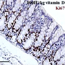
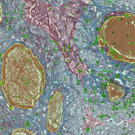
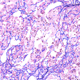
In the present experiment, 4 μm sections were taken and Ki67 stain applied (1:400, Novus Biologicals, USA) with hematoxylin (DAKO, Austria) as counter stain, on a Ventana Discovery XT autostainer (Ventana, USA), after pretreating with cell conditioner 1. The detection system used was the rabbit OmniMap (Ventana). The automated TissueFAXS PLUS system (TissueGnostics, Austria) was used to acquire whole section images.
To evaluate the sections, at least 10 intact crypts per colon were identified and the percentage of Ki67-positive cells per crypt was quantified in a blinded fashion. Care was taken to include both the ascending and descending colon.
Discussion
While research seems to indicate a role for calcium and vitamin D in preventing, modifying, or reversing neoplastic changes in colonic mucosa, the current experiment is meant to show the effect of a diet with high vitamin D content on proliferation (reduced), differentiation (increased) and apoptosis (increased), along with simultaneous upregulation of the expression of CaSR in the mouse colon.
This mechanistic model may show that molecular cross talk at the level of the calcium and vitamin D occurs directly in the colon via the CaSR, and that this is central to the anti-neoplastic effects of this combination. The gene expression and functionality of CaSR is proportional in turn to the preventive impact of active 1,25-D3 on CRC.
Prior experiments demonstrated a downregulation of the vitamin D catabolic enzyme1,25-dihydroxyvitamin D3 24-hydroxylase (CYP24A1) in the colon of mice who had higher dietary intakes of calcium. On the other hand, a low calcium intake caused colonic crypts to show increased proliferation and low apoptosis rates.
The current experiment was performed in vivo and demonstrated that a high vitamin D intake had much higher CaSR expression, decreased proliferation, and increased differentiation and apoptosis in normal colonic mucosa. The conclusion therefore was that CaSR could well link the cross talk between calcium and vitamin D, which means that their efficiency as combined chemoprophylaxis of CRC depends upon its activation and regulation by this receptor.
This year TissueGnostics (TG) turns 20! Thanks to the support of our TG product users, collaborators, distributors, and the worldwide TissueGnostics teams, TG has been able to achieve many significant milestones and has enjoyed 20 impactful years full of research, growth, and success. For this reason, TG has put together the 2023 Center of Excellence awards to celebrate and acknowledge our most impactful academic partnerships.
We are excited to finally announce our first round of Center of Excellence awardees, and to also take this opportunity to thank them again for their many years of partnership, collaborative projects and activities, and of course, publications!
Our first round takes place on the continent where it all began, Europe! However, it wouldn’t be a TissueGnostics Center of Excellence if it didn´t go global. These awards are just the beginning of many more to come, so make sure to stay connected for future updates.
Titel
StrataQuest
TissueGnostics GmbH
Taborstraße 10/2/8
1020 Vienna
Austria
Office: +43 1 216 11 90
E-mail: This email address is being protected from spambots. You need JavaScript enabled to view it.
Legal register: Handelsgericht Wien, FN 234341w
Member of the Austrian Chamber of Commerce
VAT Identification Number: ATU57176905
Website grafic and design: MATERN Creativbüro https://www.creativbuero.at
Website technique and implementation: Bettina K. Lechner, www.newhouse.at
From the very beginning TissueGnostics developed its products in close cooperation with scientists and research facilities from all over the world to ensure the effectiveness of TissueGnostics systems in everyday laboratory life. Together with Academic and Industrial Partners all over the world TissueGnostics faces the challenges of ever-changing research requirements.
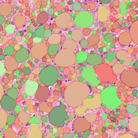
The Adipocyte APP was designed from the ground up to be run in routine by staff untrained in image analysis and image processing. The picture set is loaded into StrataQuest and the results are calculated. The algorithm automatically eliminates adipocytes on the borders from the analysis. Minor membrane tears, inevitable in working with adipose tissue sections, are automatically closed by the analysis algorithm. Larger sections of missing membrane and cell membranes collapsed into the lumina (as seen on the original image) can be drawn in, respectively out using simple manual painting tools. If this needs to be done the analysis merely has to be updated. Results can then be exported as in CSV and Excel format.
Adipocyte APP results:


TissueFAXSplus is a microscope-based cell analysis system for cells in cryocut-, paraffin-sections and/or TMAs. It consists of the software modules “TissueFAXS” combined with either “TissueQUEST” & “HistoQUEST” or with “StrataQUEST” and is used for acquisition of images in the fluorescence and/or brightfield mode, for counting the number of positive and negative cells and for quantification of staining intensities. TissueFAXSplus is used for the standardization of tissue analysis in combination with immunohistochemical and immunofluorescence staining.
TissueFAXSplus does not give any direct diagnosis and there is the possibility that the tissue specimen does not provide enough information for a proper analysis and/or diagnosis. The cell analysis system only measures cellular parameters. Such measurement parameters must in any case be reviewed and validated by a qualified human professional with profound knowledge of the cell analysis system, who has received special training in the operation of cell analysis methods. The cell analysis system can under no circumstances make a decision on the therapy of patients.
The results of the analysis are purely statistical values. Users must re-evaluate images and likelihood of the statistical data. Pure interpretation of statistical data is not allowed.
Notes:
- TissueFAXS FLUO is similar to TissueFAXSplus but is for fluorescence samples only and must not be used with brightfield/immunohistochemical samples. TissueFAXS FLUO consists of the software modules “TissueFAXS” as well as “TissueQUEST” and/or “StrataQUEST FLUO”.
- TissueFAXS HISTO is similar to TissueFAXSplus but is for brightfield/immunohistochenical samples only and must not be used with fluorescence samples. TissueFAXS HISTO consists of the software modules “TissueFAXS” as well as “HistoQUEST” and/or “StrataQUEST HISTO”.
- TissueFAXS 200 and TissueFAXS SL are similar to TissueFAXSplus but for batch scanning of up to 200/120 brightfield/immunohistochenical slides. TissueFAXS 200 / TissueFAXS SL consists of the software modules “TissueFAXS 200” / “TissueFAXS SL” as well as “HistoQUEST” and/or “TissueQuest” and/or “StrataQUEST”.
- TissueFAXS SPECTRA is similar to TissueFAXSplus but is for multispectral imaging of fluorescence samples only. Scanning of brightfield/immunohistochenical samples on TissueFAXS SPECTRA is possible with the color camera, if any, but MUST NOT / CANNOT be done with the multi-spectral camera. TissueFAXS SPECTRA consists of the software modules “TissueFAXS SPECTRA” as well as “TissueQUEST” and/or “HistoQuest” and/or “StrataQUEST”.
Using the unmixing procedure must be done with care as tissue-derived autofluorescence may interfere with the algorithm and cause inconclusive or even faulty output.
Selecting and correctly assigning proper reference spectra is critical for the accuracy of the unmixing result and is the sole responsibility of the user. Selecting wrong reference spectra and/or assigning any reference spectra inappropriately will cause inconclusive / faulty output and thus generate false measurement results!
- StrataQuest provides a software tool for machine learning for the automatic classification of tissue structures, including detection of tumor areas in tissue sections. The results generated by the software have to be verified by a qualified human professional in any case (negative as well as positive results). In case the software does not detect specific histological structures and/or tumor cells, a human professional has to verify this result by other means, as it is possible that certain biological patterns and/or special types of (cancer) cells may not be detected by the automatic detection function. Measurement errors may also occur due to the fact that the cell environment in which the software has to operate is highly variable.
We point out to the fact that the following circumstances/factors might influence and/or impair the result of the analysis to the level where the result rendered might be inconclusive or even faulty:
These factors (as well as the capability of the human professional performing the validation of the test results) lie solely within the responsibility of the user of the software. TissueGnostics does not take any responsibility for test results that are influenced by one of the above mentioned factors.
Each and any product shall be used only after training performed by TissueGnostics or authorized distributors of TissueGnostics. A list of authorized distributors is available on the TissueGnostics website.
https://tissuegnostics.com/global-network/distributors
The TissueFAXS system and software is FOR RESEARCH USE ONLY.
By using only parts of the system, changing system components (hardware or software) or using the TissueFAXSplus instrument in any other than the intended way it was designed for, the CE declaration becomes invalid.
Cytometry encompasses several methods for investigating cells, such as their count, cell cycle state, phenotype, morphology, size etc. ...

We have a lot of exciting plans to share for our big birthday, but we also have many important milestones to reflect on and remember...

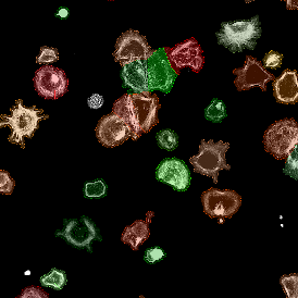
The IHC Immune Status in Situ App uses the AI classifier to segment tissue into morphological entities such as tumor, stroma, and lymphocyte clusters. It further identifies single cells ...

Explore our online database of reference publications to find tout how tissue cytometry can elevate your research
Explore our vast catalog of analysis solutions in our Strataquest Appcenter and get inspired by the variety of applications.


Read about tissue cytometry's potential in understanding the complexities of cell populations in the tissue environment

TissueGnostics GmbH
Taborstraße 10/2/8
1020 Vienna, Austria
+43 1 216 11 90
This email address is being protected from spambots. You need JavaScript enabled to view it.
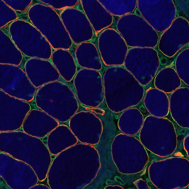
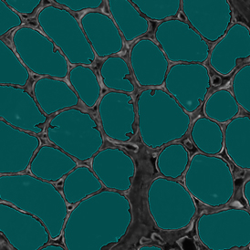
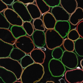
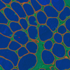
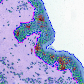
The IF Skeletal Muscle App allows the segmentation of skeletal muscle tissue sections into muscle fibers and connective tissue based on specific IF staining. Outcome parameters are provided, such as the number of muscle fibers and area of the total tissue, muscle fibers, and connective tissue.
Image: Courtesy of Stefania Petrini, Bambino Gesù Children’s Hospital, Rome
App Category 2
The IHC Adipocyte App quantifies adipocytes and their lumen in adequate HE samples. The App automatically mends small rips in adipocyte membranes and eliminates cell membrane artifacts in adipocyte lumina and lumina on sample borders. The App also outputs area measurements for all detected adipocyte lumina.
App Category 1
The IF 2 App provides single cell-based co-expression analysis for two IF markers. It segments cells into their nucleus, perinuclear area, and/or cytoplasm. Each segmented cell compartment is measured for up to 20 intensity, statistic, and morphometric parameters that can be displayed in and exported into scattergrams and histograms.
App Category 1
The IF 3 App provides single cell-based co-expression analysis for three IF markers. It segments cells into their nucleus, perinuclear area, and/or cytoplasm. Each segmented cell compartment is measured for up to 20 intensity, statistic, and morphometric parameters that can be displayed in and exported into scattergrams and histograms.
App Category 1
The IF 4 App provides single cell-based co-expression analysis for four IF markers. It segments cells into their nucleus, perinuclear area, and/or cytoplasm. Each segmented cell compartment is measured for up to 20 intensity, statistic, and morphometric parameters that can be displayed in and exported into scattergrams and histograms.
App Category 2
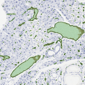
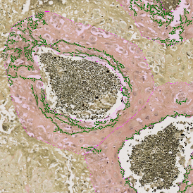
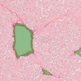
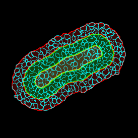
The IF Glomeruli App provides the detection of tissue, cells, and glomeruli stained by a specific marker. It segments the cells into their nucleus and/or cytoplasm and determines the cellular phenotype of specific IF-stained cell populations. The detected cells can be classified as being either inside or outside the glomeruli within certain distances (distance ranges are definable). For each cell, the spatial information and up to 20 intensity, statistics, and morphometric parameters are measured. The data can be displayed in diagrams and exported.
App Category 3
The IF Hi-Plex 50 App combines and analyses images of the same IF-stained tissue section, acquired up to 50 times with different markers. The App enables the detection of the cellular phenotypes of specific IF-stained cell populations. It segments cells into their nucleus, perinuclear area, and/or cytoplasm. Each segmented cell compartment is measured for up to 20 intensity, statistic, and morphometric parameters that can be displayed in and exported into scattergrams and histograms.
App Category 3
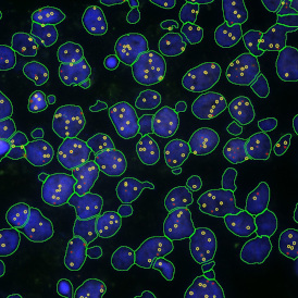
The IF Dots App provides dot detection per cell within the cell compartments for up to four markers in a sample (e.g., FISH, RNA, oil droplets). Each segmented cell compartment is measured for up to 20 intensity, statistic, and morphometric parameters. Dot measurement parameters are provided per cell compartment (e.g., nucleus, cytoplasm) and per dot and include count, mean intensity, total dot area, the sum of intensity.
App Category 2
The IF Immune Status in Situ App provides a phenotypic characterization of immune cells in reference to detected metastructures (e.g., tumors, glands) and measures the distance of detected cellular objects to the metastructure boundary (within and/or outside). Distance ranges are also definable. Each segmented cell compartment is measured for up to 20 intensity, statistic, and morphometric parameters, as well as the distance of each cell to the areas boundary.
App Category 3
The IF Skin Morphology App provides tissue detection, including the segmentation of epidermis and dermis, based on specific IF staining. It segments cells into their nucleus, perinuclear area, and/or cytoplasm and determines the cellular phenotype of specific IF-stained cell populations. The detected cells can be classified and visualized as being within or outside detected structures (epidermis and dermis). Each segmented cell compartment is measured for up to 20 intensity, statistic, and morphometric parameters that can then be displayed in diagrams and exported.
App Category 3
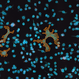
The IF Granuloma App detects granulomas based on nuclear structure analysis and an adequate IF staining (e.g., CD11c, CD68). The App measures the number and area of Granulomas and their density. Each segmented cell compartment is measured for up to 20 intensity, statistic, and morphometric parameters.
App Category 2
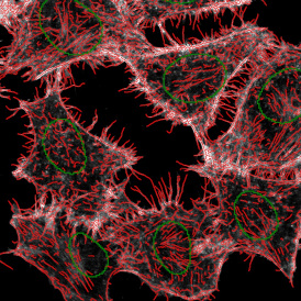
The IF Cytoskeleton App detects cytoskeletal structures based on a specific stain. The cell cytoplasm can be detected using other stains. Data can also be exported, including the number of cytoskeletal filaments inside and outside the cell and on the cell membrane, filament length, and total filament area.
App Category 3
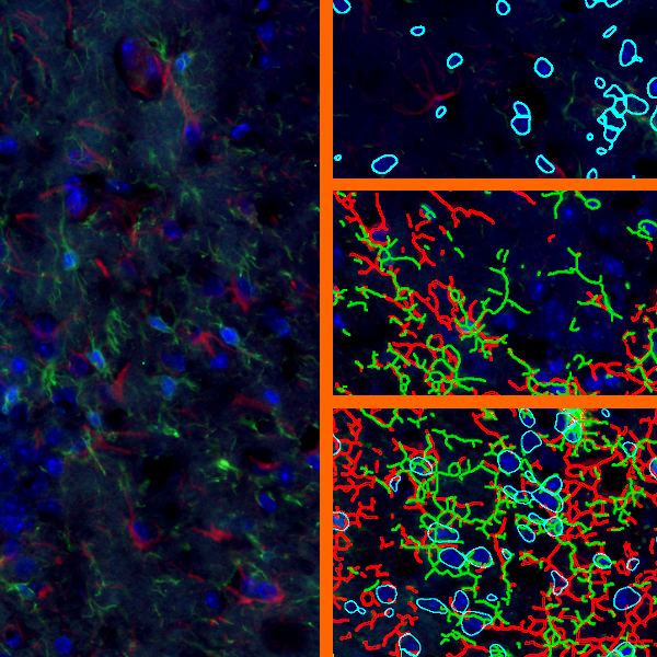
The IF Glial Cells App allows the detection of astrocytes and microglia based on specific IF staining. The measurements assessed by the App include the number of astrocytes and microglia and the number of branches of each cell (short and long attached).
App Category 3
The IHC 2 App unmixes two markers (e.g., chromogen and counterstain) in an IHC or HC digital slide and segments single cells into their nucleus, perinuclear area, and/or cytoplasm. Each segmented cell compartment is measured for up to 20 intensity, statistic, and morphometric parameters that can be displayed in and exported into scattergrams and histograms.
App Category 1
The IHC 3 App unmixes three markers (e.g., two chromogens and a counterstain) in an IHC or HC digital slide and segments single cells into their nucleus, perinuclear area, and/or cytoplasm. Each segmented cell compartment is measured for up to 20 intensity, statistic, and morphometric parameters that can be displayed in and exported into scattergrams and histograms.
App Category 2
The IHC Macrophages App detects macrophages based on adequately stained IHC samples. The App can be combined with area detection and distance range algorithms to determine the distance of Langerhans cells from the border of the epidermis inside and outside the epidermis (see above example). Each segmented cell compartment is measured for up to 20 parameters, as is the distance of each cell to the boundary.
App Category 3
The IHC Tumor-Stroma App combines the segmentation of tumor and stroma (based on the morphology) and the detection of specifically stained cell populations. It segments the cells into their nucleus, perinuclear area, and/or cytoplasm. Each segmented cell compartment in tumor and/or stroma is measured for up to 20 intensity, statistic, and morphometric parameters that can be displayed in and exported into scattergrams and histograms.
App Category 2
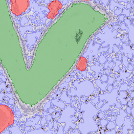
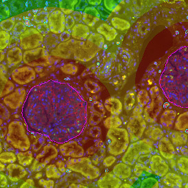
The Pulmo App segments nuclei and the metastructure components of the lung, including tissue, bronchioles, blood vessels, and alveoles. Each segmented metastructure is measured for up to 20 morphometric parameters.
App Category 3
The IHC Angio App detects blood vessels based on appropriate stains (e.g., CD31) and measures overall vessel and lumen areas. The vessel detection can be set to close open stained vessel walls and connect separated vessel sections within a definable distance. As well as vessel number, density, and areas, the App also outputs endothelium and lumina areas.
App Category 2
The IHC Meta Cells App combines the detection of IHC/HC stained metastructures (e.g., Langerhans islets, Tumor - Stroma) with single-cell detection (segmentation of cells into nucleus, perinuclear area, and/or cytoplasm). Detected cells can be classified and visualized as being within or outside detected metastructures. Each detected area and cell compartment is measured for up to 20 intensity, statistic, and morphometric parameters.
App Category 2
The RNA Scope App enables the detection of nuclei based on appropriate staining and dot detection per cell within nucleus and/or cytoplasm for one dot marker in CISH and SISH experiments . Each segmented cell compartment is measured for up to 20 intensity, statistic, and morphometric parameters. Dot parameters are provided per cell and per dot and include count, mean intensity, total dot area and the sum of intensity.
App Category 2
The Tumor Foci App allows for the detection of the whole tissue and, more importantly, tumor foci based on nuclear structure analysis, mainly on HE staining. The number, area, and density of tumor foci are measured.
App Category 1
The IHC Membrane App unmixes up to three markers in an IHC or HC digital slide and segments cells into nucleus, perinuclear area, and/or cytoplasm, as well as into membrane (e.g., HER2/neu). Each segmented cell compartment is measured for up to 20 intensity, statistic, and morphometric parameters. Three more parameters are measured for membrane intensity and angle of staining. All parameters are displayed in scattergrams and histograms and can be exported.
App Category 2
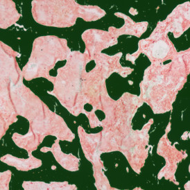
The Bone Tissue Analysis Goldner App allows for the detection of mineralized bone tissue and osteoid based on Goldner-stained bone tissue sections. The App assesses parameters such as BV (bone volume), TV (trabecular bone volume), OV (osteoid volume), OV/BV, OV/TV, OS (osteoid surface), BS (bone surface length), and the mean of osteoid width and thickness.
App Category 2
The Bone Mineralization APP separates Safranin O-stained bone tissue into its morphological substructures (cartilage, mineralized cartilage, bone marrow, and mineralized bone). Measurements assessed with this App include TV (trabecular bone volume), BV (total bone volume), MCV (mineralized cartilage), CV (cartilage volume), and bone marrow (BM).
App Category 3
The Bone Tissue Analysis Von Kossa App allows for the detection of mineralized bone tissue based on Von Kossa stained bone tissue sections. The App provides parameters such as TV (trabecular bone volume), BV (bone volume), BS (bone surface), BV/TV, BS/BV, Tb.N (trabecular number), tb.Th (trabecular thickness), and Tb.Sp (trabecular separation).
App Category 2
The IF Leishmaniasis App detects intracellular Leishmania parasites and segments them in the detected host cells. The number of parasites per cell is determined, and living and dead parasites can be distinguished (live/dead assays). The App outputs 20 intensity, statistic, and morphometric parameters for each segmented cell compartment per marker, as well as the number, mean intensity, sum of intensity, and size of parasites.
App Category 2
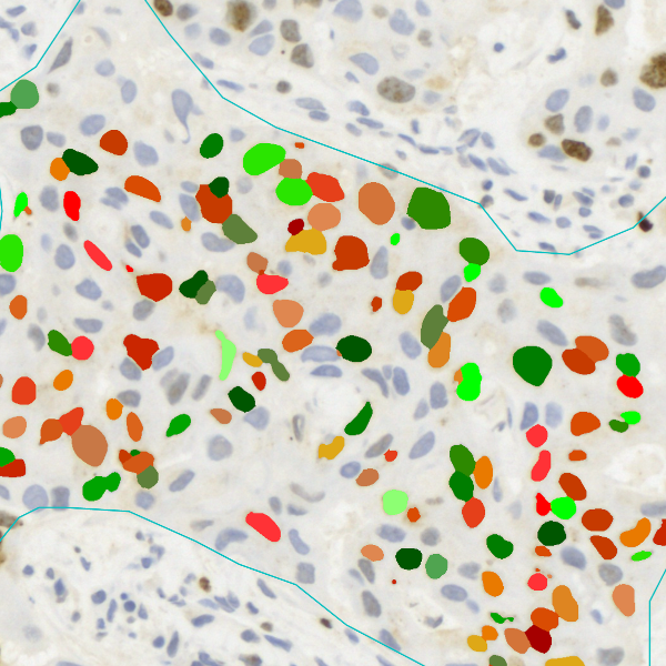
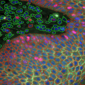
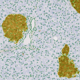
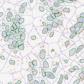
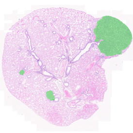
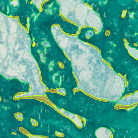
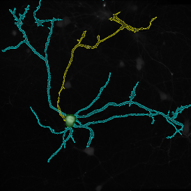
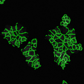
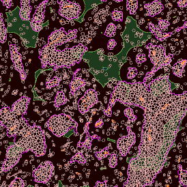
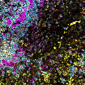
The IF Cellular Microenvironment App allows to determine the cellular phenotype of specific IF-stained cell populations and establishes their spatial relationship between each other and their neighboring cells/cell populations, including those with metastructures (e.g., blood vessels, tumors) in their vicinity. It is especially suited for proximity and infiltration analyses.
App Category 4
The IF Neurite App identifies neuronal cells and cell clusters and their neurites/dendrites. It quantifies the number of neurites/dendrites branching out from a specific neuron, identifies branch points, and exports total neurite/dendrite area, total neurite/dendrite length, average neurite/dendrite thickness, the number of branch points, and the number of endpoints.
App Category 3
The IF Pyknotic Nuclei App provides tissue detection and cell segmentation in combination with the detection of pyknotic nuclei (defined as completely condensed, round, high-intensity nuclei) based on nuclei staining. Additionally, the App allows for the determination of the cellular phenotype of specific IF-stained cell populations and dot detection. It segments the detected cells into nucleus, perinuclear area, and/or cytoplasm. The App provides parameters such as the number, mean intensity, and percentage of specific cell populations (including cells containing pyknotic nuclei). It also outputs dot parameters per segmented cell and/or dot, including count, mean intensity, total dot area, and sum of intensity.
App Category 3
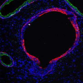
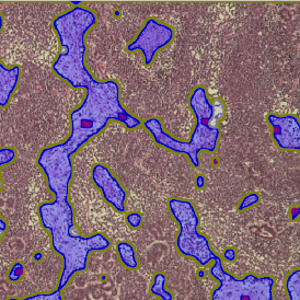
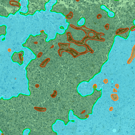
The IF Tumor Vascularization App provides tissue detection, including the separation of tumor tissue and tumor stroma (healthy tissue). Additionally, it detects blood vessels based on appropriate stains (e.g., CD31) and measures the number, area, and density of these blood vessels. The vessel detection also can be set to close open stained vessel walls and to connect separated vessel sections within a definable distance.
App Category 3
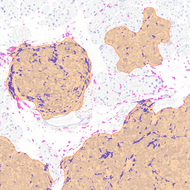
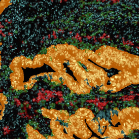
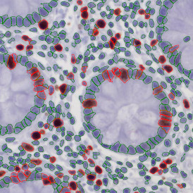
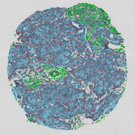
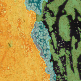
The IHC Tumor-Macrophages App provides tissue detection, including the separation of tumor and healthy tissue. It detects macrophages based on specific staining (e.g., CD68) and outputs the area of macrophages within tumor and healthy tissue.
Image: Courtesy of Dr. Patrick Michl, Dr. Maren Egidi, and Dr. Heidi Griesmann, Universitätsklinikum Halle (Saale).
App Category 3
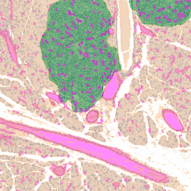
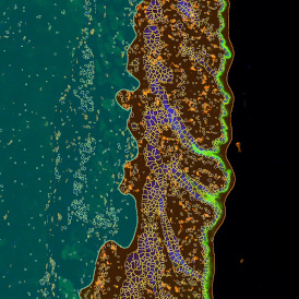
The IHC Tumor Vascularization App provides tissue detection, including the separation of tumor tissue and tumor stroma (healthy tissue). Additionally, it detects blood vessels based on appropriate stains (e.g., CD31) and measures the number and area of these blood vessels. The vessel detection can also be programmed to close open stained vessel walls and to connect separated vessel sections within a definable distance. The App outputs the number, density, and areas of vessels within both tumor and healthy tissue.
Image: Courtesy of Dr. Patrick Michl, Dr. Maren Egidi, and Dr. Heidi Griesmann, Universitätsklinikum Halle (Saale).
App Category 3
The IHC Angio Trichome App detects blood vessels based on trichome staining and measures the overall vessel and lumen area. Furthermore, it detects specifically IHC-stained single cell populations and establishes their spatial relationship to the detected blood vessels. The App outputs number and vessel density, vessel wall thickness and areas of vessels, endothelium and lumina, the number of IHC stained cells, proximity measurement, etc.
App Category 3
The RNA Scope+ App provides detection of nuclei based on appropriate staining and dot detection per cell within the nucleus and/or cytoplasm for two dot markers in CISH and SISH experiments. Each segmented cell compartment is measured for up to 20 intensity, statistic, and morphometric parameters. It also outputs dot parameters per segmented cell and/or dot, including count, mean intensity, total dot area, and sum of intensity.
App Category 2
The IHC Angio Elastin App detects blood vessels in Verhoeffs van Geison-stained samples, elastin, and collagen. The outputs include the number and area of vessels, elastin, and collagen within a definable distance to the vessel.
App Category 2
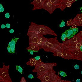
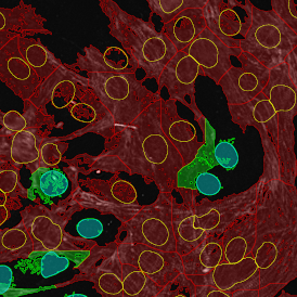
The IF Cardio Cell Culture App provides cell segmentation, detection of cardiomyocytes (based on appropriate staining, e.g., Troponin Red), fibroblasts within cultured cardio cells, plus one additional marker. The App outputs parameters such as the number of cardiomyocytes, fibroblasts, and marker-positive cardiomyocytes and fibroblasts.
Image: Courtesy of Agatha Ribeiro da Silva, Prof. Jose E. Krieger (Heart Institute, University Sao Paulo)
App Category 3
The IF Cardio Cell Culture Dot App provides cell segmentation and detection of cardiomyocytes (based on an appropriate stain, e.g., Troponin Red) and fibroblasts within cultured cardio cells, plus one dot marker (CISH, FISH). The App outputs parameters such as the number of cardiomyocytes and fibroblasts. It also outputs the number of dot-positive cardiomyocytes and fibroblasts and the number, area (μm²), and mean intensity of dots per cell.
Image: Courtesy of Agatha Ribeiro da Silva, Prof. Jose E. Krieger (Heart Institute, University Sao Paulo).
App Category 3
The IF Dendrites & Axons App identifies neuronal cells, their dendrites and axon, based on appropriate markers. It quantifies the number of dendrites branching out from a specific neuron. The App provides the total number of dendrites per neuron, including the length of these dendrites and their axons.
Image: Courtesy of Thomas Bastian, Ph.D., University of Minnesota.
App Category 3
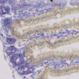
The IHC Small Intestine - Dots App provides nuclei segmentation and detection of tissue and villi based on nuclei staining (crypts need to be defined manually). Furthermore, it allows dot detection for one dot markers (CISH, RNAScope, SISH) within villi and crypt areas. Dot parameters are provided for villi and crypts and for dots and include count, mean intensity, total dot area, the sum of intensity.
App Category 2
The Lipid Droplets App quantifies lipid droplets within H&E stained tissues (e.g., liver). The App automatically mends small rips in liver droplet membranes and eliminates cell membrane artifacts, including lumina on sample borders. The App also outputs area and number measurements for all detected lipid droplets.
App Category 1
The Angio Sirius Red App detects collagen and blood vessels based on Sirius Red staining. The app outputs the area of Sirius Red stained collagen and the number of detected vessels.
App Category 2
The IHC Angio Diameter App detects blood vessels based on appropriate stains (e.g., CD31). The App outputs vessel area, number, density, and blood vessel diameter.
App Category 2
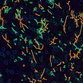
The IF Cultured Cells & Substructures App detects cells based on nuclei staining, as well as one dot marker (FISH, CISH experiments) and cytoskeletal structures based on a specific stain. It outputs the number of detected cells, the number and intensity of dots per cell, and the density of cytoskeletal filaments.
App Category 3
The IF Membrane App detects nuclei and segments the cells into different cellular compartments, including membrane, nuclei, and cytoplasm. It also detects one additional marker (e.g., HER2/neu). Each segmented cell compartment is measured for different parameters, such as staining intensity, stained area, and the number/percentage of marker-positive cells within the detected cellular compartments. Three more parameters are measured for the membrane, including membrane area, membrane length, and the angle of staining.
App Category 2
The IF Spheroids App allows a comprehensive analysis of spheroids (as well as organoids and embryoid bodies). It automatically identifies the spheroids and cells based on the nuclei staining and analyzes two additional IF markers. It segments the cells into different cellular compartments, including membrane, nuclei, and cytosol, and further measures the marker expression for each compartment. It can also measure dot markers (if available). It establishes proximity distances for the cells detected within the spheroids, bringing the IF-stained cell populations into spatial context.
App Category 3
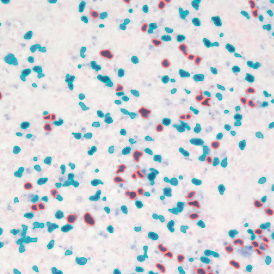
The IHC Immune Status in Situ App uses the AI classifier to segment tissue into morphological entities such as tumor, stroma, and lymphocyte clusters. It further identifies single cells based on nuclei staining (hematoxylin), detects immune cells based on appropriated stains (CD45, CD3, CD20, etc.), and measures the distance of detected cells to the metastructure boundary. The App can also define distance ranges through outputting parameters, including the area of the detected morphological entities and the number/percentage of lymphocytes detected within the tissue entities, as well as in certain proximities.
App Category 3
The Wilms Tumor App is based on the AI Classifier and allows for the segmentation of H&E stained Wilms tumor tissues into tumor, stroma, and blood vessels. It outputs the area (µm2) of the segmented tissue entities.
App Category 1
The IHC Necrotic Tumor App segments tumor tissues into tumor, necrotic tissue, and stroma using the AI Classifier. Furthermore, it identifies single cells as well as one additional cellular marker (e.g., neutrophils). It outputs the area of tumor, necrotic tissue, and stroma and measures the number and percentage of neutrophils within the morphological entities.
App Category 3
The IHC Necrotic Tumor Angio App can segment tumor tissues into tumor, necrotic tissue, and blood vessels using the AI Classifier. It outputs the area of tumor, necrotic tissue, and blood vessels, as well as the number and percentage of blood vessels in total and within the two morphological entities.
App Category 3
The IF Rods & Cones in Retina App detects the rods and cones based on specific staining. It outputs the number, density, and length of detected structures, as well as the number, percentage, and density of marker-stained rods and cones.
App Category 2
The IF Tumor Foci Angio App identifies single cells and segments tissues into tumor foci and blood vessels based on appropriate markers. It applies proximity maps to identify nuclei close to blood vessels. It measures the number of nuclei located within a certain distance relative to blood vessels, the number of nuclei in the different morphological entities, and the area of these morphological entities.
App Category 3
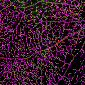
The IF Retinal Vasculature App detects blood vessels in retinal tissue based on appropriate stains (e.g., CD31). The App outputs vessel area, density, and length.
App Category 2
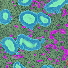
The Endometrium HE App allows for the segmentation of hematoxylin and eosin-stained endometrium tissues into their morphological entities (glands, stroma, and blood vessels). The measurements provided by the App include the area of glands, stroma, and blood vessels.
App Category 2
The IHC Extracellular Filament App detects nuclei and extracellular filaments stained with specific markers. It outputs the number of nuclei, total filaments area, and the length of these filaments.
App Category 2
The IF Cellular Contact App allows for the determination of the cellular phenotype of specific IF-stained cell populations and establishes the cellular contacts to their neighboring cells (the number of markers is technically unlimited). If needed, the App provides a separation of nuclei in tissues with high cellular densities. The App outputs parameters such as staining intensity per marker and morphometric parameters for each segmented cell/cell compartment, as well as the number and percentage of cells of different phenotypes in direct contact.
Images: courtesy of Naoki Kaneko/Shiv Pillai (PI), Ragon Institute of MGH, MIT and Harvard, Boston, MA USA.
App Category 3
The App was used in a CELL publication, read more
The EBER-ISH App analyzes tissue samples stained by EBER-ISH probes (EBV-encoded RNA in-situ hybridization). These probes visualize the Epstein-Barr virus (EBV) EBER RNA. First, the App identifies nuclei, then detects EBER-ISH positive nuclei. The measurements provided by the App include the number of detected cells and the number, density, and percentage of EBER-ISH positive cells.
App Category 1
The IF Cell Culture - Osteoclast App allows for the segmentation of nuclei, the identification of cultured multinucleated osteoclasts (stained by a specific marker), and the quantification of one or two additional markers. Outputs include the number of detected cells, osteoclasts, and nuclei per osteoclast, the area of osteoclasts, and the intensity of markers within the osteoclasts.
App Category 2
read more about Automated Detection and Characterization of Osteoclasts in Microscopic Images
Privacy Notice
This website uses YouTube videos. In order to access the video, please acknowledge that it will be loaded from a YouTube server. Personal data may be transmitted to YouTube. You'll find further information here.