TissueFAXS Fluo
UPGRADES
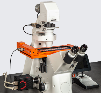
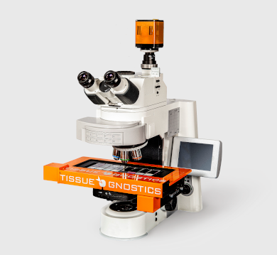
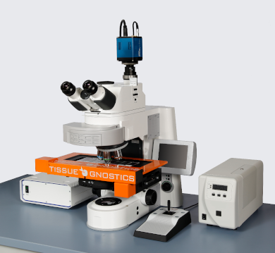
All imaging systems are modular and upgradable. Every system can be customized to offer the following capabilities: brightfield scanning, widefield fluorescence, confocal, high-throughput and multispectral. To exemplify TissueFAXS Fluo can be ugraded to
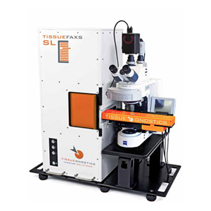
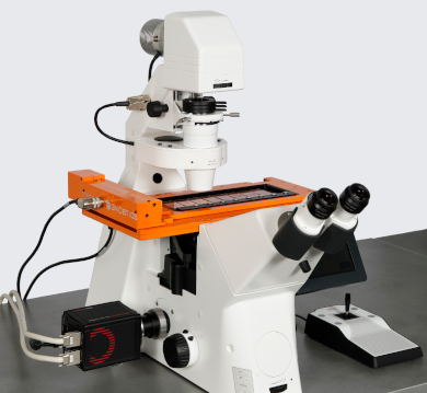
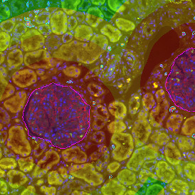
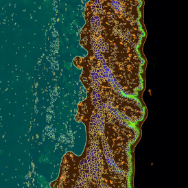
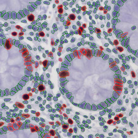
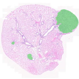
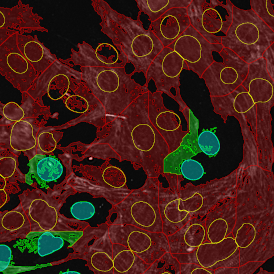
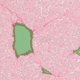
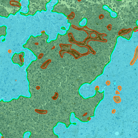
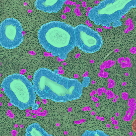
- TissueFAXS PLUS (image on the left)
- TissueFAXS Q
- TissueFAXS SPECTRA
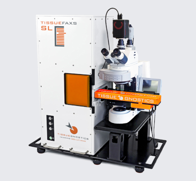
- TissueFAXS SL
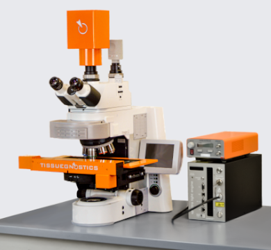
TissueFAXS Q
TECHNOLOGY
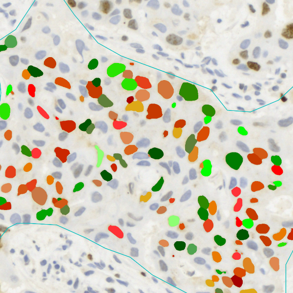
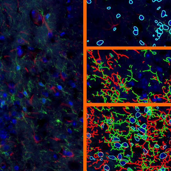
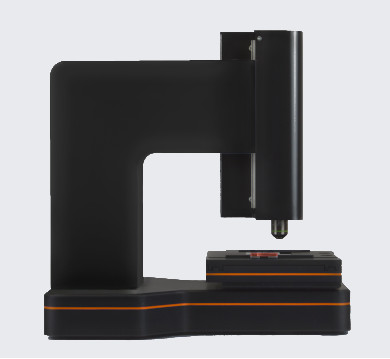
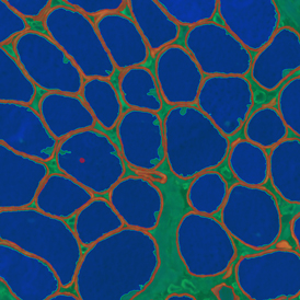
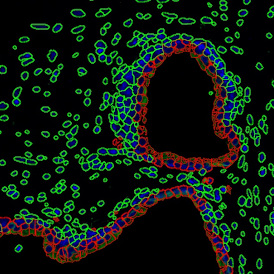
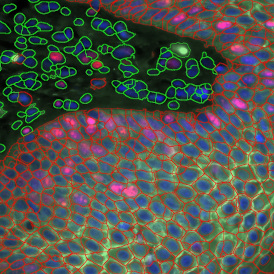
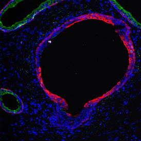
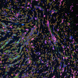
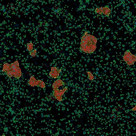
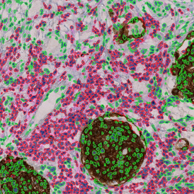
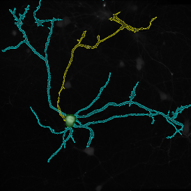
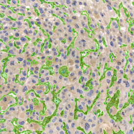
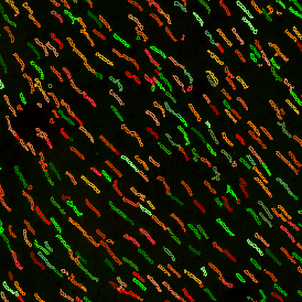
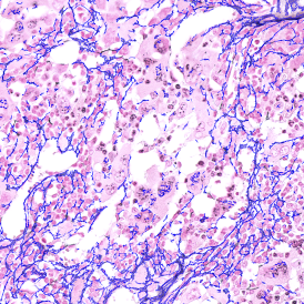
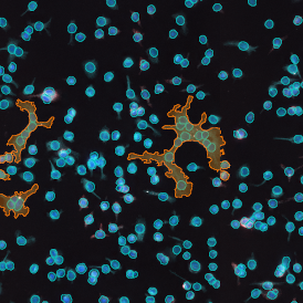
The TissueFAXS SL Q performance can scan a complete slice of mouse brain with a size of 73.3 mm 2 with four channels and a 13 step Z-stack in 1.5 hours with 20x magnification (see image on the right). This is possible through the interplay of the components of the TissueFAXS SL Q.
TissueFAXS Q Spinning Disk unit
The Spinning Disc Unit of the TissueFAXS Q is a versatile, high tech, yet affordable Spinning Disc Device. The unit is mounted on the front port of the systems quadrocular phototube and supported externally for stability. It features:
- Widefield (with fast disk out motorized function) or Confocal (single or double pattern) Double Pinhole Pattern Spinning Disk mode, on the same physical disk
- Single Pinhole Pattern Spinning Disk for large Field of View Motorized Dichroic Five positions Filter Wheel
- Standard Eight positions Motorized Emission Wheel
- Plug-in spinning disk
- Fast spinning disk 15,000 RPM disk rotation speed
- Extraction tools for easy insertion and removal of both dichroic filter and emission filter
- Detector focal plane easy focusing without moving the detector
TissueFAXS Q System cameras and light sources
The standard camera used on the TissueFAXS Q is a superior alternative (speed, quality) to the standard TissueFAXS Fluo camera.
The light engines are used for high power fluorescence illumination. These illumination systems combine very high output, necessary for confocality, with very short switching times (<10 ns rise and fall times between bands). Highly intense and constant white light is provided by an VIS-LED with high 25,000 hours of illuminant life.
TissueFAXS SPECTRA SL
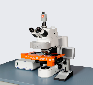
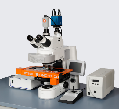
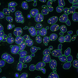
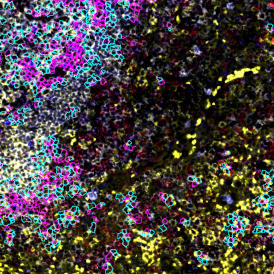
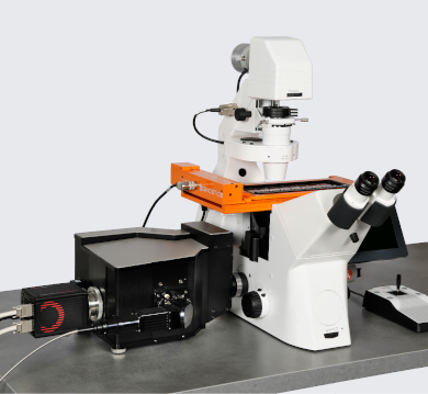
All imaging systems are modular and upgradable. Every system can be customized to offer the following capabilities: brightfield scanning, widefield fluorescence, confocal, multispectral as well as high-throughput. To exemplify TissueFAXS SPECTRA can be upgraded to the high-throughput TissueFAXS SPECTRA SL (image on the left side),
- high-throughput scanning in multispectral fluorescence, widefield fluorescence as well as brightfield mode
- fully automated loading/scanning of 120 slides at a time
- three 40 slide magazines with each magazine containing 20 two slide metal clips for a total of 120 standard slides.
- alternatively 60 double sized slides
- this system also supports TMA scanning